Review
. 2020 Apr 15:11:494.
doi: 10.3389/fimmu.2020.00494. eCollection 2020.

The Immune Response to the fVIII Gene Therapy in Preclinical Models

Affiliations
Review

The Immune Response to the fVIII Gene Therapy in Preclinical Models

Seema R Patel et al. Front Immunol. .

Abstract

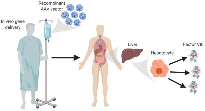
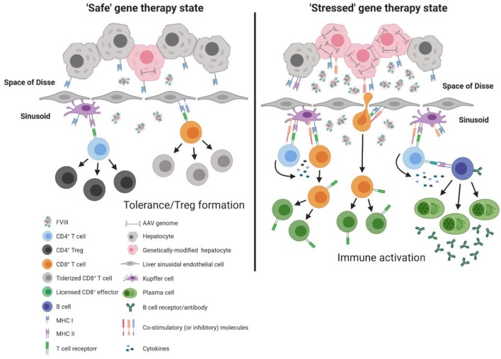
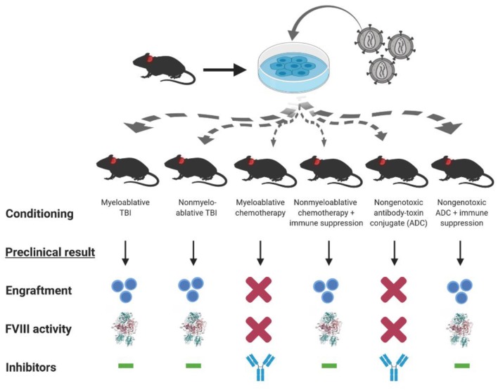
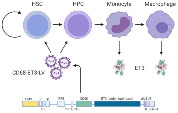
Neutralizing antibodies to factor VIII (fVIII), referred to as "inhibitors," remain the most challenging complication post-fVIII replacement therapy. Preclinical development of novel fVIII products involves studies incorporating hemophilia A (HA) and wild-type animal models. Though immunogenicity is a critical aspect of preclinical pharmacology studies, gene therapy studies tend to focus on fVIII expression levels without major consideration for immunogenicity. Therefore, little clarity exists on whether preclinical testing can be predictive of clinical immunogenicity risk. Despite this, but perhaps due to the potential for transformative benefits, clinical gene therapy trials have progressed rapidly. In more than two decades, no inhibitors have been observed. However, all trials are conducted in previously treated patients without a history of inhibitors. The current review thus focuses on our understanding of preclinical immunogenicity for HA gene therapy candidates and the potential indication for inhibitor treatment, with a focus on product- and platform-specific determinants, including fVIII transgene sequence composition and tissue/vector biodistribution. Currently, the two leading clinical gene therapy vectors are adeno-associated viral (AAV) and lentiviral (LV) vectors. For HA applications, AAV vectors are liver-tropic and employ synthetic, high-expressing, liver-specific promoters. Factors including vector serotype and biodistribution, transcriptional regulatory elements, transgene sequence, dosing, liver immunoprivilege, and host immune status may contribute to tipping the scale between immunogenicity and tolerance. Many of these factors can also be important in delivery of LV-fVIII gene therapy, especially when delivered intravenously for liver-directed fVIII expression. However, ex vivo LV-fVIII targeting and transplantation of hematopoietic stem and progenitor cells (HSPC) has been demonstrated to achieve durable and curative fVIII production without inhibitor development in preclinical models. A critical variable appears to be pre-transplantation conditioning regimens that suppress and/or ablate T cells. Additionally, we and others have demonstrated the potential of LV-fVIII HSPC and liver-directed AAV-fVIII gene therapy to eradicate pre-existing inhibitors in murine and canine models of HA, respectively. Future preclinical studies will be essential to elucidate immune mechanism(s) at play in the context of gene therapy for HA, as well as strategies for preventing adverse immune responses and promoting immune tolerance even in the setting of pre-existing inhibitors.

Keywords: adeno-associated viral vectors; factor VIII (fVIII); gene therapy; hematopoietic (stem) cells; hemophilia A; inhibitors; lentiviral (LV) vector.


Figures

Figure 1
Figure 1
In vivo AAV-fVIII gene therapy. AAV-fVIII vectors selected for hepatocyte tropism and encompassing a fVIII transgene cassette under a liver-specific transcriptional promoter are infused into adult patients via peripheral vein. Once in circulation, the AAV vectors are thought to transduce primarily hepatocytes, persist episomally, and direct biosynthesis and secretion of fVIII into the bloodstream.
Figure 2
Figure 2
Model of immune response to liver directed AAV-fVIII gene therapy. The liver is a unique immunoprivileged site that, through complex interactions of an array of liver immune constituents, teeters between tolerance and inflammation. These immune populations include liver sinusoidal endothelial cells (LSECs) that line the wall of the sinusoids and are intimately associated with resident macrophages of the liver (Kupffer cells), hepatic stellate cells (Ito cells) that reside in the space of Disse between hepatocytes and LSECs, and hepatic dendritic cells that reside in the sinusoidal lumen of the liver. Under basal conditions, an array of immune constituents (e.g., Kupffer cells and LSECs) express low levels of MHC and co-stimulatory molecules as well as immunomodulatory cytokines. In the absence of cellular stress following AAV-fVIII gene therapy (“safe” gene therapy state), the local immunomodulatory milieu of the liver can suppress the activation of vector specific and fVIII reactive T cells. Moreover, expression of co-inhibitory molecules by LSECs can aid in the efficient differentiation of fVIII specific Tregs. However, a bolus infusion of AAV particles and/or overexpression of fVIII can lead to cellular stress that possesses the capacity to deviate the immune environment from immunomodulatory to pro-inflammatory. Under AAV-fVIII gene therapy mediated cellular stress (“stressed” gene therapy state), genetically modified hepatocytes can up-regulate MHC class I and co-stimulatory molecules as well as the production of pro-inflammatory cytokines. CD8+ T cell recognition of cognate antigens expressed by “stressed” hepatocytes can be activated, ultimately resulting in the cytolysis of genetically modified hepatocytes and decline in fVIII production. In addition, the pro-inflammatory milieu generated from cellular stress can promote differentiation of effector fVIII specific CD4+ T cells that can help activate fVIII specific B cells for formation of inhibitors.
Figure 3
Figure 3
Conditioning dependent outcomes of preclinical HSPC LV-fVIII gene therapy. CD34+ HSPC isolated from hemophilia A or congenic mice are genetically modified ex vivo using LV-fVIII gene therapy. Transduced cells then are infused into naïve (or preimmunized with recombinant fVIII) hemophilia A mice in the presence or absence of various myeloablative and non-myeloablative conditioning regimens that are based on clinical transplantation protocols. Of the regimens tested in the preclinical stetting, myeloablative and non-myeloablative total body irradiation (TBI), or chemotherapy plus T cell immunosuppression (anti-thymocyte globulin or co-stimulation blockade), allowed for engraftment and corrective fVIII activity levels in the absence of inhibitor formation.
Figure 4
Figure 4
Ex vivo CD68-ET3-LV CD34+ clinical gene therapy paradigm. Autologous CD34+ HSPC are isolated from subjects with hemophilia A, genetically modified ex vivo using LV encompassing a codon optimized pfVIII transgene (ET3) under the monocyte lineage restricted promoter, CD68. Genetically modified HSPCs are then infused back into the subject following non-myeloablative conditioning with immune suppression. Post-administration of the genetically-modified autologous cell product, plasma fVIII levels, vector copy number in peripheral blood, and fVIII immunity status are followed.

References

    1. Gitschier J, Wood WI, Goralka TM, Wion KL, Chen EY, Eaton DH, et al. . Characterization of the human factor VIII gene. Nature. (1984) 312:326–30. 10.1038/312326a0 - DOI - PubMed
    1. Toole JJ, Knopf JL, Wozney JM, Sultzman LA, Buecker JL, Pittman DD, et al. . Molecular cloning of a cDNA encoding human antihaemophilic factor. Nature. (1984) 312:342–7. 10.1038/312342a0 - DOI - PubMed
    1. Lollar P. Pathogenic antibodies to coagulation factors. Part one: factor VIII and factor IX. J Thromb Haemost. (2004) 2:1082–95. - PubMed
    1. Brackmann HH, Gormsen J. Massive factor-VIII infusion in haemophiliac with factor-VIII inhibitor, high responder. Lancet. (1977) 2:933. 10.1016/S0140-6736(77)90871-6 - DOI - PubMed
    1. Dorner AJ, Bole DG, Kaufman RJ. The relationship of N-linked glycosylation and heavy chain-binding protein association with the secretion of glycoproteins. J Cell Biol. (1987) 105:2665–74. 10.1083/jcb.105.6.2665 - DOI - PMC - PubMed

Publication types

MeSH terms