Giant Cell Tumor of the Calcaneus
- PMID: 32351846
- PMCID: PMC7187992
- DOI: 10.7759/cureus.7467
Giant Cell Tumor of the Calcaneus
Abstract
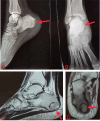
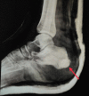
A 17-year-old female presented to us with pain and swelling in the right heel. Examination revealed the swelling to be tender, hard and fixed to the calcaneus. Radiographs showed an expansile, lytic lesion of the calcaneus with well-defined margins and no extraosseus spread. A core biopsy was done which showed multinucleated giant cells in a sea of mononuclear stromal cells, suggestive of a giant cell tumour (GCT). Curettage and filling up of the defect with bone cement was done under anaesthesia. The patient was fully ambulatory three months after the surgery. At two-year follow-up, the patient continued to be asymptomatic and radiographs revealed no signs of recurrence. It is important to note that GCT can occur in these rare sites and unusual age groups, and hence requires a good level of awareness of the surgeon and adequate preoperative workup, including biopsy, before proceeding to the definitive treatment of the lesion. Considering its potential local aggressiveness, early intervention is necessary. The patient should be kept under regular follow-up to detect any recurrence or metastasis in early stage.
Keywords: bone cementing; calcaneum; calcaneus; curettage; gct; giant cell tumor; recurrence.
Copyright © 2020, Batheja et al.
Conflict of interest statement
The authors have declared that no competing interests exist.
Figures


References
-
- Giant cell tumor of bone. Campanacci M, Baldini N, Boriani S, Sudanese A. https://www.ncbi.nlm.nih.gov/pubmed/3805057. J Bone Joint Surg. 1987;69:106–113. - PubMed
-
- Giant‐cell tumor: a study of 195 cases. Dahlin DC, Cupps RE, Johnson EW Jr. Cancer. 1970;25:1061–1070. - PubMed
-
- Giant cell tumour of foot bones-25 years experience in a tertiary care hospital. Minhas MS, Khan KM, Muzzammil M. https://www.ncbi.nlm.nih.gov/pubmed/26878540 J Pak Med Assoc. 2015;65:0. - PubMed
Publication types
LinkOut - more resources
Full Text Sources
