. 2020 Apr 8:2020:5753623.
doi: 10.1155/2020/5753623. eCollection 2020.

Sinapic Acid Promotes Browning of 3T3-L1 Adipocytes via p38 MAPK/CREB Pathway

Affiliations

Sinapic Acid Promotes Browning of 3T3-L1 Adipocytes via p38 MAPK/CREB Pathway

In-Seon Bae et al. Biomed Res Int. .

Abstract

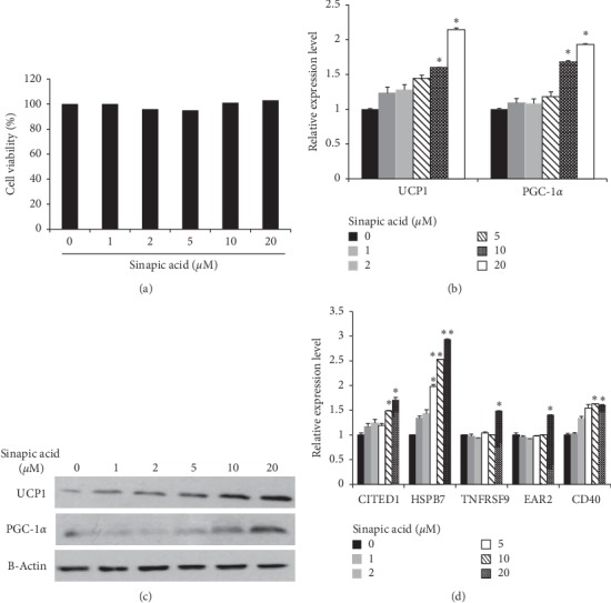
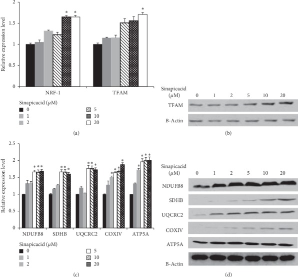
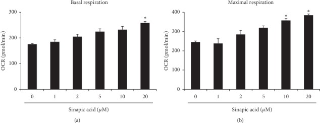
Sinapic acid is a plant-derived phenolic compound, which acts as an antioxidant, anticancer, and anti-inflammatory agent. Although sinapic acid is valuable in a variety of therapeutic applications, its role in the improvement of obesity-related metabolic disease is relatively unexplored. Brown-like adipocytes (beige adipocytes) are characterized by a high concentration of mitochondria and high expression of uncoupling protein 1 (UCP1), which has specific functions in energy expenditure and thermogenesis. This study assessed the browning effects of sinapic acid in 3T3-L1 adipocytes. We investigated the expression of beige marker genes in 3T3-L1 adipocytes treated with sinapic acid. Sinapic acid increased the expression of peroxisome proliferator-activated receptor γ coactivator-1α (PGC-1α) and UCP1. Sinapic acid also promoted mitochondrial biogenesis by dose-dependently upregulating the oxygen consumption rate and enhancing the expression of representative subunits of oxidative phosphorylation complexes. In addition, treatment with p38 mitogen-activated protein kinase (MAPK) inhibitor and cAMP response element binding (CREB) inhibitor decreased the expressions of genes associated with thermogenesis, mitochondrial biogenesis, and oxidative phosphorylation. In summary, sinapic acid initiates browning 3T3-L1 adipocytes via the p38 MAPK/CREB signaling pathway. Thus, sinapic acid may have potential therapeutic implication in obesity.


Conflict of interest statement

The authors report no conflicts of interest.

Figures

Figure 1
Figure 1
Browning of adipocytes in 3T3-L1 cells was induced by sinapic acid. (a) 3T3-L1 cells were treated with different concentrations of sinapic acid and cell viability was measured. (b, c) The expression of thermogenic genes (UCP1 and PGC-1α) was analyzed by qRT-PCR analysis (b) and western blot analysis (c) in 3T3-L1 adipocytes treated with sinapic acid. P < 0.05. (d) The relative mRNA expression levels of beige-specific genes (CITED1, HSPB7, TNFRSF9, EAR2, and CD40) were measured by qRT-PCR in 3T3-L1 cells treated with different doses of sinapic acid. P < 0.05, ∗∗P < 0.01.
Figure 2
Figure 2
Sinapic acid stimulates mitochondrial biogenesis. (a) The effect of sinapic acid on NRF-1 and TFAM expression was analyzed in sinapic acid-treated 3T3-L1 adipocytes. P < 0.05. (b) The protein level of TFAM in 3T3-L1 cells treated with sinapic acid was measured by western blot analysis. (c, d) The expression of mitochondrial biogenesis related genes (NDUFB8, SHDB, UQCRC2, COXIV, and ATA5A) in sinapic acid-treated 3T3-L1 adipocytes was detected by qRT-PCR analysis (c) and western blot analysis (d) P < 0.05.
Figure 3
Figure 3
Sinapic acid promotes mitochondrial activity. Cells were cultured in different concentrations of sinapic acid. Mitochondrial respiration of 3T3-L1 adipocytes was measured using the XF-24 Extracellular Flux Analyzer. (a) Basal respiration was analyzed before the sequential addition of complex inhibitors in the mitochondrial electron transport chain. P < 0.05. (b) Maximal respiration was assessed after the addition of carbonyl cyanide p-trifluoromethoxyphenylhydrazine as an uncoupling agent. P < 0.05.
Figure 4
Figure 4
p38 MAPK and CREB mediate the browning effect of sinapic acid in 3T3-L1 cells. (a) 3T3-L1 adipocytes were treated with sinapic acid at the indicated concentrations. The levels of p38 MAPK, CREB, AKT, and AMPK proteins were measured by western blotting. (b, c) Cells exposed to 20 μM sinapic acid were treated with the p38 MAPK inhibitor SB 203580 (b) and the CREB inhibitor 666-15 (c). The level of mitochondrial biogenesis-related genes and thermogenic genes in 3T3-L1 adipocytes were analyzed by western blot analysis.

References

    1. Giralt M., Villarroya F. White, brown, beige/brite: different adipose cells for different functions? Endocrinology. 2013;154(9):2992–3000. doi: 10.1210/en.2013-1403. - DOI - PubMed
    1. Seale P., Lazar M. A. Brown fat in humans: turning up the heat on obesity. Diabetes. 2009;58(7):1482–1484. doi: 10.2337/db09-0622. - DOI - PMC - PubMed
    1. Choe S. S., Huh J. Y., Hwang I. J., Kim J. I., Kim J. B. Adipose tissue remodeling: its role in energy metabolism and metabolic disorders. Frontiers in Endocrinology. 2016;7:p. 30. doi: 10.3389/fendo.2016.00030. - DOI - PMC - PubMed
    1. Peres Valgas da Silva C., Hernández-Saavedra D., White J., Stanford K. Cold and exercise: therapeutic tools to activate Brown adipose tissue and combat obesity. Biology. 2019;8(1):p. 9. doi: 10.3390/biology8010009. - DOI - PMC - PubMed
    1. El Hadi H., Di Vincenzo A., Vettor R., Rossato M. Food ingredients involved in white-to-brown adipose tissue conversion and in calorie burning. Frontiers in Physiology. 2019;9:p. 1954. doi: 10.3389/fphys.2018.01954. - DOI - PMC - PubMed

MeSH terms

Substances