. 2020 Apr 24:3:61.
doi: 10.1038/s41746-020-0266-y. eCollection 2020.

PENet-a scalable deep-learning model for automated diagnosis of pulmonary embolism using volumetric CT imaging

Affiliations

PENet-a scalable deep-learning model for automated diagnosis of pulmonary embolism using volumetric CT imaging

Shih-Cheng Huang et al. NPJ Digit Med. .

Erratum in

Abstract

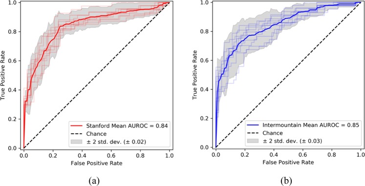
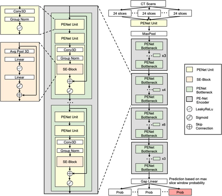
Pulmonary embolism (PE) is a life-threatening clinical problem and computed tomography pulmonary angiography (CTPA) is the gold standard for diagnosis. Prompt diagnosis and immediate treatment are critical to avoid high morbidity and mortality rates, yet PE remains among the diagnoses most frequently missed or delayed. In this study, we developed a deep learning model-PENet, to automatically detect PE on volumetric CTPA scans as an end-to-end solution for this purpose. The PENet is a 77-layer 3D convolutional neural network (CNN) pretrained on the Kinetics-600 dataset and fine-tuned on a retrospective CTPA dataset collected from a single academic institution. The PENet model performance was evaluated in detecting PE on data from two different institutions: one as a hold-out dataset from the same institution as the training data and a second collected from an external institution to evaluate model generalizability to an unrelated population dataset. PENet achieved an AUROC of 0.84 [0.82-0.87] on detecting PE on the hold out internal test set and 0.85 [0.81-0.88] on external dataset. PENet also outperformed current state-of-the-art 3D CNN models. The results represent successful application of an end-to-end 3D CNN model for the complex task of PE diagnosis without requiring computationally intensive and time consuming preprocessing and demonstrates sustained performance on data from an external institution. Our model could be applied as a triage tool to automatically identify clinically important PEs allowing for prioritization for diagnostic radiology interpretation and improved care pathways via more efficient diagnosis.

Keywords: Cardiovascular diseases; Radiography.


Conflict of interest statement

Competing interestsThe authors declare no competing interests.

Figures

Fig. 1
Fig. 1. PENet performance on independent test datasets.
Receiver operating characteristic curve (ROC) with bootstrap confidence intervals on Stanford internal test set (a) and Intermountain external test set (b).
Fig. 2
Fig. 2
(Sensitivity vs. specificity plot): Sensitivity and specificity across different operating point (probability threshold) with increment of 0.05 on the Stanford internal test set (a) and Intermountain external test (b).
Fig. 3
Fig. 3
(Class Activation Maps): Class activation map (CAM) representation of true positive (Stanford (a) and Intermountain (b), false-positive (Stanford (c) and Intermountain (d) and false-negative samples (Stanford (e) and Intermountain (f)—axial contrast enhanced CT pulmonary angiogram (left) and CAM inferred by the model overlay with the scan (right). a (Stanford test set: true positive): (left) demonstrates a non-occlusive filling defect in a left lower pulmonary artery segmental branch that is correctly localized by the model as seen in the CAM image overlay (right). b (Intermountain test set: True Positive): (left) demonstrates a non-occlusive filling defect in a left main pulmonary artery that is correctly localized by the model as seen in the CAM image overlay (right). c (Stanford test set: false positive): left) demonstrates a large left hilar node adjacent to the pulmonary artery that is incorrectly labeled as PE by the model as seen in the CAM image overlay (right). d (Intermountain test set: false positive): (left) demonstrates an enlarged unopacfied left lower lobe pulmonary vein invaded by tumor that is incorrectly labeled as PE by the model as seen in the CAM image overlay (right). e (Stanford test set: false negative): (left) Pulmonary embolism in right middle lobe segmental branch that is missed by the model as seen in the CAM image overlay (right). f (Intermountain test set: False negative): (left) Pulmonary embolism in left upper lobe segmental branch that is missed by the model as seen in the CAM image overlay (right).
Fig. 4
Fig. 4. PENet architecture used in this study.
PENet is built using four architectural units: the PENet unit, Squeeze-and-Excitation block, the PE-Net bottleneck, and the PE-Net encoder. Each building block in the network is color-coded.

References

    1. Horlander KT, Mannino DM, Leeper KV. Pulmonary embolism mortality in the United States, 1979–1998: an analysis using multiple-cause mortality data. Arch. Intern. Med. 2003;163:1711–1717. doi: 10.1001/archinte.163.14.1711. - DOI - PubMed
    1. Le Gal G, Bounameaux H. Diagnosing pulmonary embolism: running after the decreasing prevalence of cases among suspected patients. J. Thrombosis Haemost. 2004;2:1244–1246. doi: 10.1111/j.1538-7836.2004.00795.x. - DOI - PubMed
    1. Leung AN, et al. An Official American Thoracic Society/Society of Thoracic Radiology clinical practice guideline: evaluation of suspected pulmonary embolism in pregnancy. Am. J. Respir. Crit. Care Med. 2011;184:1200–1208. doi: 10.1164/rccm.201108-1575ST. - DOI - PubMed
    1. Leung AN, et al. American Thoracic Society documents: an official American Thoracic Society/Society of Thoracic Radiology clinical practice guideline—evaluation of suspected pulmonary embolism in pregnancy. Radiology. 2012;262:635–646. doi: 10.1148/radiol.11114045. - DOI - PubMed
    1. Yavas US, Calisir C, Ozkan IR. The Interobserver Agreement between Residents and Experienced Radiologists for Detecting Pulmonary Embolism and DVT with Using CT Pulmonary Angiography and Indirect CT Venography. Korean J. Radiol. 2008;9:498–502. doi: 10.3348/kjr.2008.9.6.498. - DOI - PMC - PubMed