. 2020 Jun;24(12):6822-6832.
doi: 10.1111/jcmm.15336. Epub 2020 Apr 30.

Trametinib potentiates TRAIL-induced apoptosis via FBW7-dependent Mcl-1 degradation in colorectal cancer cells

Affiliations

Trametinib potentiates TRAIL-induced apoptosis via FBW7-dependent Mcl-1 degradation in colorectal cancer cells

Lin Lin et al. J Cell Mol Med. 2020 Jun.

Abstract

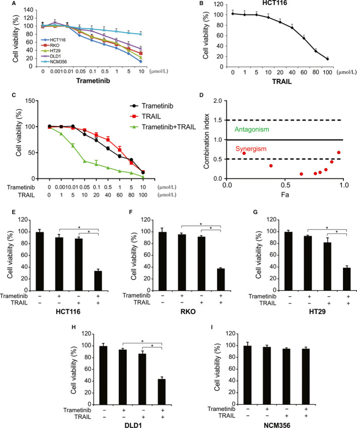
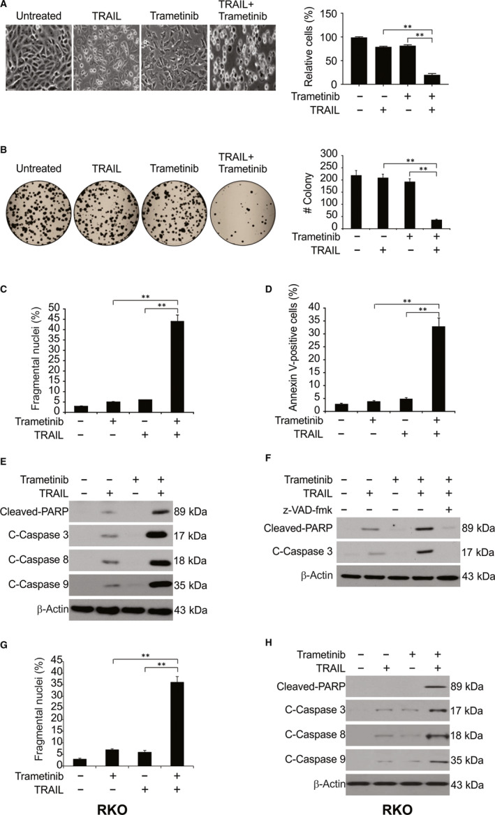
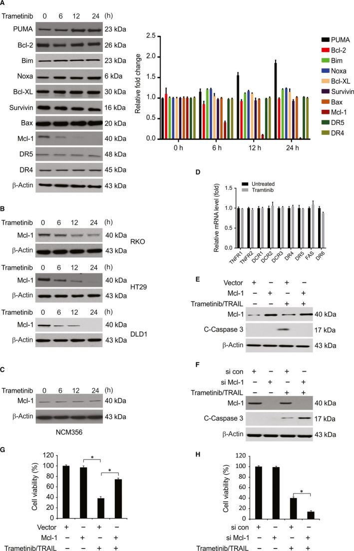
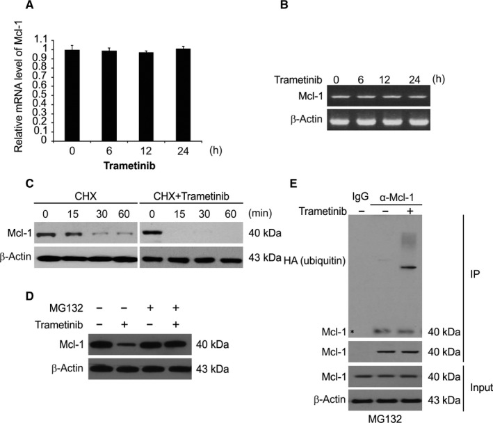
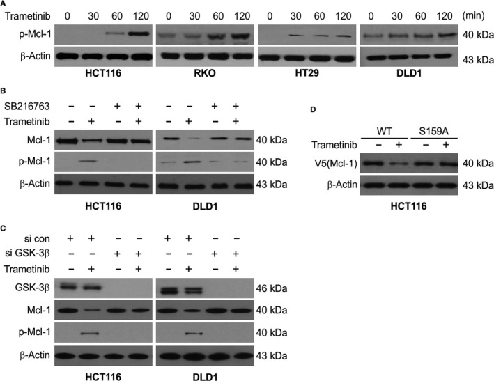
Trametinib is a MEK1/2 inhibitor and exerts anticancer activity against a variety of cancers. However, the effect of Trametinib on colorectal cancer (CRC) is not well understood. In the current study, our results demonstrate the ability of sub-toxic doses of Trametinib to enhance TRAIL-mediated apoptosis in CRC cells. Our findings also indicate that Trametinib and TRAIL activate caspase-dependent apoptosis in CRC cells. Moreover, Mcl-1 overexpression can reduce apoptosis in CRC cells treated with Trametinib with or without TRAIL. We further demonstrate that Trametinib degrades Mcl-1 through the proteasome pathway. In addition, GSK-3β phosphorylates Mcl-1 at S159 and promotes Mcl-1 degradation. The E3 ligase FBW7, known to polyubiquitinate Mcl-1, is involved in Trametinib-induced Mcl-1 degradation. Taken together, these results provide the first evidence that Trametinib enhances TRAIL-mediated apoptosis through FBW7-dependent Mcl-1 ubiquitination and degradation.

Keywords: Mcl-1; TRAIL; Trametinib; apoptosis; degradation.


Conflict of interest statement

The authors have declared that no conflicts of interest exist.

Figures

Figure 1
Figure 1
Trametinib promotes TRAIL‐induced cytotoxicity of human CRC cell lines. A, Indicated cells were treated with various concentrations of Trametinib for 72 h. Cell growth was analysed by MTT. B, HCT116 cells were treated with various concentrations of TRAIL for 72 h. Cell growth was analysed by MTT. C, HCT116 cells were treated with Trametinib and TRAIL at indicated concentration for 72 h. Cell growth was analysed by MTT. D, Combination index is shown for HCT116 cells. Fa, fraction affected. E‐I, Indicated cells were incubated in the presence or absence of TRAIL (10 ng/mL) and/or Trametinib (0.1 μmol/L) for 72 h. Cell growth was analysed by MTT. Results in (E)‐(I) were expressed as means ± SD of three independent experiments. *, P < 0.05
Figure 2
Figure 2
Trametinib sensitizes TRAIL‐induced apoptosis in CRC cells. A, HCT116 cells were treated with 0.1 μmol/L Trametinib, 10 ng/mL TRAIL or their combination 24 h. Cell morphology was examined under a light microscope. Attached cells were counted. B, HCT116 cells plated in six‐well cell culture plates were treated with 0.1 μmol/L Trametinib, 10 ng/mL TRAIL, or their combination for 24 h. After 14 days, the plates were stained for cell colonies with crystal violet dye, and photographs of colonies taken using a digital camera. C, HCT116 cells were treated with 0.1 μmol/L Trametinib, 10 ng/mL TRAIL or their combination for 24 h. Apoptosis was analysed by a nuclear fragmentation assay. D, HCT116 cells were treated with 0.1 μmol/L Trametinib, 10 ng/mL TRAIL, or their combination for 24 h. Apoptosis was analysed by Annexin V/PI staining followed by flow cytometry. E, HCT116 cells were treated with 0.1 μmol/L Trametinib, 10 ng/mL TRAIL, or their combination for 24 h. Indicated proteins were analysed by Western blotting. F, HCT116 cells pre‐treated with 10 μmol/L z‐VAD‐fmk for 1 h were treated with 0.1 μmol/L Trametinib, 10 ng/mL TRAIL, or their combination for 24 h. Indicated proteins were analysed by Western blotting. G, RKO cells were treated with 0.1 μmol/L Trametinib, 10 ng/mL TRAIL or their combination for 24 h. Apoptosis was analysed by a nuclear fragmentation assay. H, RKO cells were treated with 0.1 μmol/L Trametinib, 10 ng/mL TRAIL or their combination for 24 h. Indicated proteins were analysed by Western blotting. Results in (B), (C), (D) and (G) were expressed as means ± SD of three independent experiments. **, P < 0.01
Figure 3
Figure 3
Trametinib‐induced Mcl‐1 down‐regulation is associated with the induction of TRAIL‐mediated apoptosis. A, HCT116 cells were treated with 0.1 μmol/L Trametinib at indicated time point. Indicated protein level was determined by Western blotting and normalized to β‐actin. B, Indicated cell lines were treated with 0.1 μmol/L Trametinib at indicated time point. Mcl‐1 level was analysed by Western blotting. C, NCM356 cells were treated with 0.1 μmol/L Trametinib at indicated time point. Mcl‐1 level was analysed by Western blotting. D, HCT116 cells were treated with 0.1 μmol/L Trametinib for 24 h. Relative mRNA levels of indicated gene were analysed by real‐time PCR. E, HCT116 cells transfected with Mcl‐1 were treated with the combination of 0.1 μmol/L Trametinib and 10 ng/mL TRAIL for 24 h. Cleaved caspase 3 was analysed by Western blotting. F, HCT116 cells transfected with si control or si Mcl‐1 were treated with the combination of 0.1 μmol/L Trametinib and 10 ng/mL TRAIL for 24 h. Cleaved caspase 3 was analysed by Western blotting. G, HCT116 cells transfected with Mcl‐1 were treated with the combination of 0.1 μmol/L Trametinib and 10 ng/mL TRAIL for 72 h. Cell growth was analysed by MTT. H, HCT116 cells transfected with si control or si Mcl‐1 were treated with the combination of 0.1 μmol/L Trametinib and 10 ng/mL TRAIL for 72 h. Cell growth was analysed by MTT. Results in (D), (G) and (H) were expressed as means ± SD of three independent experiments. *, P < 0.05
Figure 4
Figure 4
Trametinib promotes Mcl‐1 degradation and ubiquitination. A, HCT116 cells were treated with 0.1 μmol/L Trametinib at indicated time point. Mcl‐1 mRNA level was analysed by RT‐qPCR, with β‐actin as a control. B, HCT116 cells were treated with 0.1 μmol/L Trametinib at indicated time point. Total RNA was extracted, and Mcl‐1 mRNA expression was analysed by semiquantitative reverse transcription PCR, followed by gel electrophoresis. β‐actin was used as a control. C, HCT116 cells were treated with or without Trametinib in the presence of cyclohexamide (CHX) (10 μg/mL) for the indicated time periods. The Mcl‐1 protein level was determined by Western blotting. D, Trametinib‐treated cells were treated with or without MG132 and subjected to Western blotting. E, HCT116 cells transfected with HA‐ubiquitin and pre‐treated with 5 μmol/L MG132 for 30 min were treated 0.1 μmol/L Trametinib for 4 h. IP was performed to pull down Mcl‐1, followed by Western blotting of indicated proteins
Figure 5
Figure 5
FBW7 is required for Trametinib‐induced Mcl‐1 degradation and ubiquitination. A, HCT116 cells were treated with 0.1 μmol/L Trametinib for 24 h. IP was performed to pull down Mcl‐1, followed by Western blotting of indicated proteins. B, Parental and FBW7 knockdown HCT116 cells transfected with HA‐ubiquitin and pre‐treated with 5 μmol/L MG132 for 30 min were treated 0.1 μmol/L Trametinib for 4 h. IP was performed to pull down Mcl‐1, followed by Western blotting of indicated proteins
Figure 6
Figure 6
GSK3β mediates Trametinib‐induced Mcl‐1 phosphorylation and degradation. A, Indicated cell lines were treated with 0.1 μmol/L Trametinib at indicated time point. Phosphorylation of Mcl‐1 was analysed by Western blotting. B, HCT116 and DLD1 cells were pre‐treated with 1 μmol/L SB216763 for 1 h and then treated with 1 μmol/L Trametinib for an additional 2 h. Indicated protein level was determined by Western blotting. C, HCT116 and DLD1 cells transfected with si control or GSK3β siRNA were treated with 0.1 μmol/L Trametinib for an additional 2 h. Indicated protein level was determined by Western blotting. D, HCT116 cells transfected with WT or S159A Mcl‐1. After 24 h, the cells were treated with 0.1 μmol/L Trametinib for an additional 2 h. Indicated protein level was determined by Western blotting

References

    1. De Rosa M, Pace U, Rega D, et al. Genetics, diagnosis and management of colorectal cancer (Review). Oncol Rep. 2015;34:1087‐1096. - PMC - PubMed
    1. Tong J, Zheng X, Tan X, et al. Mcl‐1 phosphorylation without degradation mediates sensitivity to HDAC inhibitors by liberating BH3‐only proteins. Can Res. 2018;78:4704‐4715. - PMC - PubMed
    1. Modest DP, Pant S, Sartore‐Bianchi A. Treatment sequencing in metastatic colorectal cancer. Eur J Cancer. 2019;109:70‐83. - PubMed
    1. Yaeger R, Corcoran RB. Targeting alterations in the RAF‐MEK pathway. Cancer Discov. 2019;9:329‐341. - PMC - PubMed
    1. Oliveira AF, Bretes L, Furtado I. Review of PD‐1/PD‐L1 inhibitors in metastatic dMMR/MSI‐H colorectal cancer. Front Oncol. 2019;9:396. - PMC - PubMed

Publication types

MeSH terms