. 2020 Apr 30;15(4):e0231977.
doi: 10.1371/journal.pone.0231977. eCollection 2020.

Reduced cellular binding affinity has profoundly different impacts on the spread of distinct poxviruses

Affiliations

Reduced cellular binding affinity has profoundly different impacts on the spread of distinct poxviruses

Erica B Flores et al. PLoS One. .

Abstract

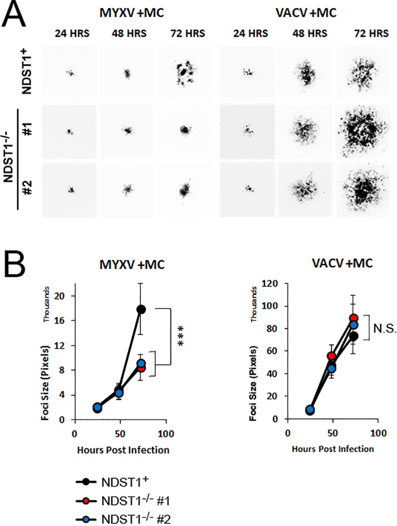
Poxviruses are large enveloped viruses that replicate exclusively in the cytoplasm. Like all viruses, their replication cycle begins with virion adsorption to the cell surface. Unlike most other viral families, however, no unique poxviral receptor has ever been identified. In the absence of a unique receptor, poxviruses are instead thought to adhere to the cell surface primarily through electrostatic interactions between the positively charged viral envelope proteins and the negatively charged sulfate groups on cellular glycosaminoglycans (GAGs). While these negatively charged GAGs are an integral part of all eukaryotic membranes, their specific expression and sulfation patterns differ between cell types. Critically, while poxviral binding has been extensively studied using virally centered genetic strategies, the impact of cell-intrinsic changes to GAG charge has never been examined. Here we show that loss of heparin sulfation, accomplished by deleting the enzyme N-Deacetylase and N-Sulfotransferase-1 (NDST1) which is essential for GAG sulfation, significantly reduces the binding affinity of both vaccinia and myxoma viruses to the cell surface. Strikingly, however, while this lowered binding affinity inhibits the subsequent spread of myxoma virus, it actually enhances the overall spread of vaccinia by generating more diffuse regions of infection. These data indicate that cell-intrinsic GAG sulfation plays a major role in poxviral infection, however, this role varies significantly between different members of the poxviridae.


Conflict of interest statement

The authors have declared that no competing interests exist.

Figures

Fig 1
Fig 1. Generation of cells deficient in sulfation of cell surface heparin chains.
(A) PCR analysis of NDST1 mRNA expression in NDST1+ and NDST1 deficient cell lines. Bar graph shows quantitative data from real-time PCR analysis. Qualitative agarose gel is shown for visual purposes. (B) Western Blot analysis was performed using the indicated antibodies to detect expression of NDST1. Expression of actin is shown as a loading control. Data is representative of two independent experiments. (C) Phase contrast images of NDST1+ and deficient cells depicting general overall cellular morphology. (D) Cell growth of both NDST1+ and deficient cells measured over a 96-hour period. Data is representative of two independent experiments. (E) Sulfation of cell surface heparin measured via flow cytometry utilizing an antibody that recognizes the sulfated 10E4 epitope on HS chains. The mean fluorescent intensity (MFI) values were then normalized to NDST+ values to show the relative change in the NDST-/- cell lines. Data is representative of three independent experiments. Statistical significance was determined using Students T-Test. *** = p<0.001.
Fig 2
Fig 2. Loss of sulfation reduces binding of both MYXV and VACV virions.
(A) Histograms depicting binding of fluorescent viral virions to the cell surface. Cells were incubated on ice with Cy-5 labeled MYXV or VACV at 4 particles per cell and washed with cold PBS to remove residual virus. Cells were resuspended in 2% PFA and levels of Cy-5-labeled virus bound to the cell membrane was measured via flow cytometry. Data shown is representative of two independent experiments each done in triplicate. (B) Relative MFI values of Cy-5 (viral binding). MFI values were normalized to NDST+ to determine the relative change in NDST-/- cell lines. Statistical MFI calculations are a summation of the two independent experiments each conducted in triplicate. Statistical significance was determined using non-parametric Mann-Whitney test. ** = p<0.01, * = p<0.05.
Fig 3
Fig 3. Loss of sulfation reduces initial infection of both MYXV and VACV.
(A) Number of GFP+ cells found 24 hours after infection with either MYXV or VACV at multiple MOI’s. Data shown is the summation of two independent experiments each conducted in triplicate. (B) Fluorescent images of cells infected for 24 hours with MYXV at an MOI of 1, 3, and 9 shown as an example.
Fig 4
Fig 4. Loss of sulfation delays intracellular poxviral replication.
(A) Kinetic analysis of GFP expression after initial infection of NDST1+ or deficient cells. NDST1+ or deficient B16/F10 cells were infected with either MYXV or VACV and GFP expression measured every two hours using flow cytometry. Data presented is representative of two independent experiments. (B) Abundance of MYXV or VACV genomes measured using qPCR. Data presented is representative of three independent experiments for both viruses each analyzed in triplicate and is presented as the change in cycle threshold value (ΔCT) from 2 hour baseline. (C) Production of new infection viral progeny in either NDST1+ or deficient cells measured using standard single step growth curve analysis. Data presented is a summation of two (VACV) or three (MYXV) independent experiments each conducted in triplicate. Statistical significance was determined using Students T-Test (* = p<0.05).
Fig 5
Fig 5. Binding affinity has inverse impacts on the spread of different poxviruses.
(A) Images of individual GFP+ foci formed in either NDST1+ or deficient cells taken 24, 48, or 72 hours post infection with the indicated virus. (B) Quantitation of individual foci size. Data shown is representative of four independent experiments, where a total of >60 foci per cell/virus type were measured. (C) Up-close image of foci showing the different concentration of infected cells in the core. (D) Quantitation of total GFP signal in individual foci from experiments above. Data shown represents average GFP expression from >15 foci measured across three experiments. Statistical significance was determined using Students T-Test (*** = p<0.001).
Fig 6
Fig 6. Increased spread seen during low affinity VACV infections is mediated by secreted virions.
(A) Images of individual GFP+ foci formed in either NDST1+ or deficient cells covered with methyl cellulose. Images were taken 24, 48, or 72 hours post infection with the indicated virus. (B) Quantitation of individual foci size. Data is representative from at least three independent experiments. Statistical significance was determined using Students T-Test. *** = p<0.001.

References

    1. Knipe DM, Howley PM. 2013. Fields virology, 6th ed. Wolters Kluwer/Lippincott Williams & Wilkins Health, Philadelphia, PA.
    1. Esko JD, Selleck SB. 2002. Order out of chaos: assembly of ligand binding sites in heparan sulfate. Annu Rev Biochem 71:435–71. 10.1146/annurev.biochem.71.110601.135458 - DOI - PubMed
    1. Kwon MJ, Jang B, Yi JY, Han IO, Oh ES. 2012. Syndecans play dual roles as cell adhesion receptors and docking receptors. FEBS Lett 586:2207–11. 10.1016/j.febslet.2012.05.037 - DOI - PubMed
    1. Lindahl U, Kjellen L. 2013. Pathophysiology of heparan sulphate: many diseases, few drugs. J Intern Med 273:555–71. 10.1111/joim.12061 - DOI - PubMed
    1. Whitelock JM, Iozzo RV. 2005. Heparan sulfate: a complex polymer charged with biological activity. Chem Rev 105:2745–64. 10.1021/cr010213m - DOI - PubMed

Publication types