Review
. 2020 May;43(5):326-342.
doi: 10.1016/j.tins.2020.03.005. Epub 2020 Apr 15.

Prenatal Origins of ASD: The When, What, and How of ASD Development

Affiliations
Review

Prenatal Origins of ASD: The When, What, and How of ASD Development

Eric Courchesne et al. Trends Neurosci. 2020 May.

Abstract

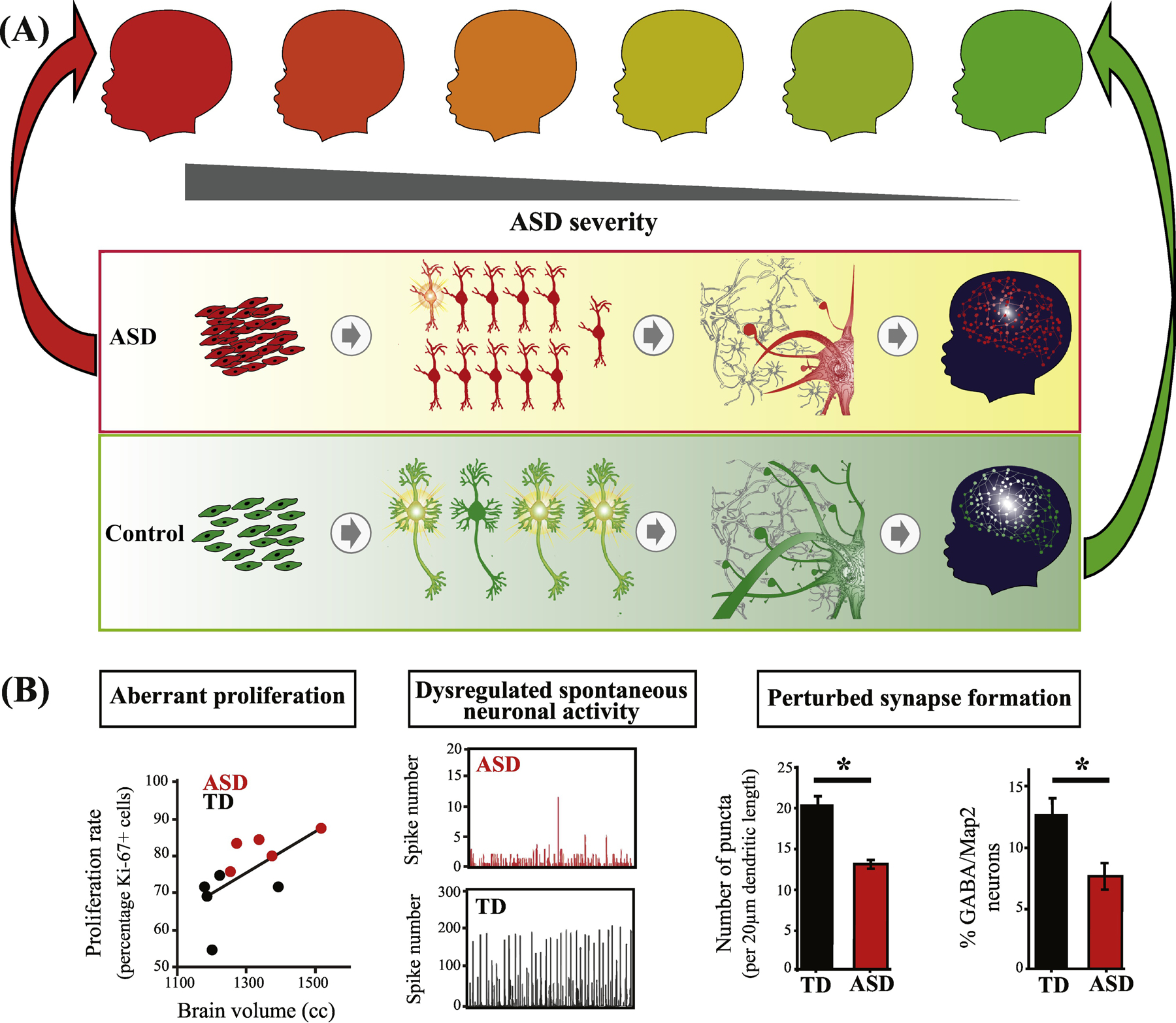
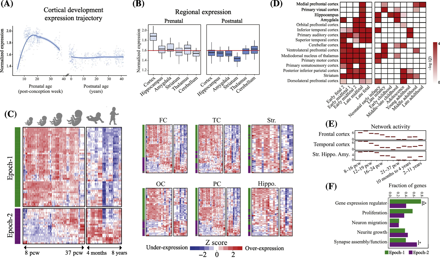
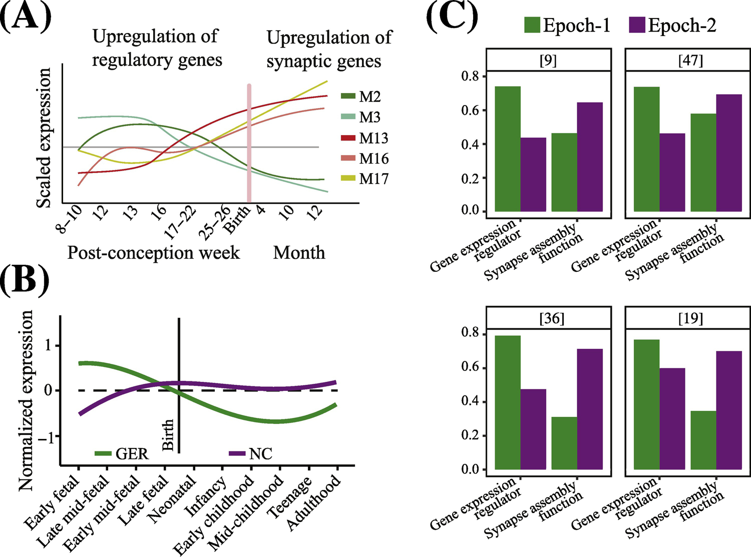
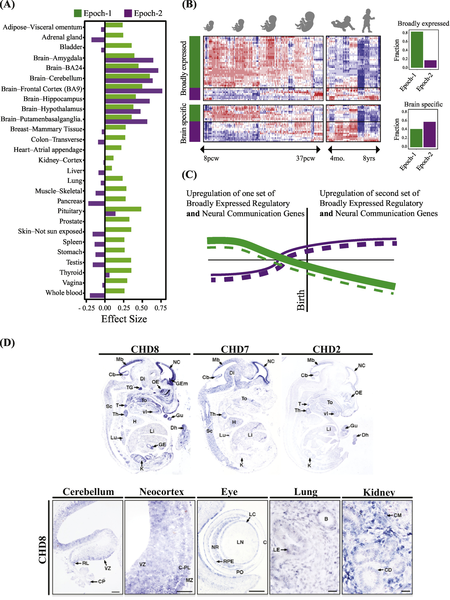
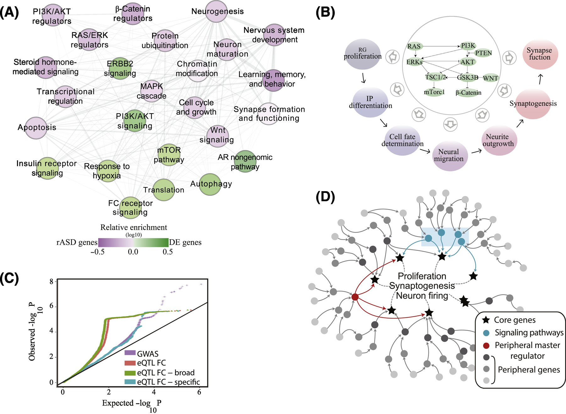
Autism spectrum disorder (ASD) is a largely heritable, multistage prenatal disorder that impacts a child's ability to perceive and react to social information. Most ASD risk genes are expressed prenatally in many ASD-relevant brain regions and fall into two categories: broadly expressed regulatory genes that are expressed in the brain and other organs, and brain-specific genes. In trimesters one to three (Epoch-1), one set of broadly expressed (the majority) and brain-specific risk genes disrupts cell proliferation, neurogenesis, migration, and cell fate, while in trimester three and early postnatally (Epoch-2) another set (the majority being brain specific) disrupts neurite outgrowth, synaptogenesis, and the 'wiring' of the cortex. A proposed model is that upstream, highly interconnected regulatory ASD gene mutations disrupt transcriptional programs or signaling pathways resulting in dysregulation of downstream processes such as proliferation, neurogenesis, synaptogenesis, and neural activity. Dysregulation of signaling pathways is correlated with ASD social symptom severity. Since the majority of ASD risk genes are broadly expressed, many ASD individuals may benefit by being treated as having a broader medical disorder. An important future direction is the noninvasive study of ASD cell biology.

Keywords: autism; brain specific; broadly expressed; gene; prenatal; proliferation; regulatory; synapse.


Figures

Figure 1.
Figure 1.. ASD is a progressive multi-stage disorder of prenatal and early postnatal development.
(A) ASD involves multiple stages of prenatal cortical maldevelopment as compared with typical development (from[9]). ASD begins with aberrant cortex formation that involves disruption of cell cycle processes and excess cell proliferation (left panel); neuronal maturation, neurite outgrowth and spontaneous neural activity (next panel); and continues with disruption of later prenatal and postnatal synaptogenesis and neural network expansion (third panel); and “wiring” of cortex and coordinated neural circuits activity (right panel)(from[9]). Upper series of child icons from red to green, illustrates the hypothesis that ASD outcome severity (darker red) may be related to severity of dysregulation in some or all of these prenatal and postnatal stages. (B) In one study, ASD subject-derived iPS cell (iPSC) models show that abnormal increases in cell proliferation are correlated with each child’s early brain overgrowth (left panel). iPSC-derived ASD neurons also show reduced spontaneous neuronal activity (middle panel) and deviant synapse formation (right panel) (from[20]). See Box 1 for more details.
Figure 2.
Figure 2.. ASD genetics uniformly implicate prenatal beginnings in multiple brain regions and multiple functional processes.
(A-B) Genes implicated in ASD by a recent GWAS study show strong expression during prenatal corticogenesis but not during postnatal cortex development (from[32]). (C) Neocortex expression pattern of 69 hcASD genes during brain development. 94% of 69 hcASD risk genes express at prenatal ages across multiple brain regions (from[9]). The heatmaps also show there are two major groups of hcASD genes: Epoch-1 (green bar in heatmaps) consisting of 68% of hcASD genes and Epoch-2 (purple bar in heatmaps) consisting of 32% (from[9]). FC: Frontal cortex; OC: Occipital cortex; PC: Parietal cortex; TC: Temporal cortex; Hippo: Hippocampus; Str: Striatum. (D) Similar to hcASD genes, an extended set of ASD risk genes show peak expression at prenatal ages across multiple brain regions (from[17]). (E) Co-expression activity of genes perturbed in leukocytes of individuals with ASD implicate prenatal neurodevelopmental stage across frontal and temporal cortices and the striatum, hippocampus and amygdala (Str.Hippo.Amy)(from[51]). (F) hcASD genes are highly pleotropic and are involved in multiple stages of prenatal neural development. Epoch-1 (green bars) is heavily dominated by regulatory hcASD genes (79% of Epoch-1 genes) that can disrupt proliferation, neurogenesis and other early stages of cortical formation, while Epoch-2 (purple bars) is dominated by brain-specific hcASD genes (>60% of Epoch-2 genes) that may disrupt late prenatal and postnatal synaptogenesis and “wiring” of cortex. Percentages across functional clusters add up to more than 100% because the majority of hcASD gene mutations affect more than one developmental process.
Figure 3.
Figure 3.. Dysregulation of regulatory ASD genes dominate the earliest prenatal stages of ASD.
The identification of critical developmental periods and changes in pathobiological molecular mechanisms in ASD is of paramount importance. (A) In one approach, researchers first cluster genes according to co-expression patterns and next examine their enrichment for ASD risk genes (from[14]). M2 and M3 = modules enriched in gene expression regulator ASD risk genes; M13, M16 and M17 = modules enriched in synaptogenesis and synapse functioning ASD risk genes. (B) In a second approach, researchers make an a priori selection of genes, modules, functions or ontologies of interest from among all possible ones, and then analyze those selected for age-related changes in expression. A recent example is shown in this panel (from[36]). GER = gene expression regulator ASD risk genes; NC = neural communication ASD risk genes. Using such a priori selection of regulatory and synapse-relevant genes shows that ASD genes that are regulators normally upregulate expression as early as 1st and 2nd trimesters and then downregulate in perinatal and postnatal life, and the opposite pattern for synapse/neural communication genes, namely downregulation in early prenatal life followed by upregulation in later prenatal and postnatal life. Note that mutations in these ASD risk genes would therefore result in their dysregulation during these critical prenatal periods and disruption of these brain development processes. This approach has led some to view this as evidence of two largely non-overlapping and non-interacting types of genes, mechanisms and processes. (C) Bar graphs of hcASD genes from four recent studies[9, 19, 36, 47]. List of hcASD genes from each study were manually annotated by literature search (see Supplementary Table S-1). hcASD genes from each study were assigned to Epoch-1 or Epoch-2 based on their correlation with the neurodevelopmental time stages considering the time point as an ordinal variable. In all four studies, regulatory and synapse-relevant hcASD genes are present in both Epoch-1 and Epoch-2. In each study, Epoch-1 has a greater percentage of regulatory hcASD genes than does Epoch-2, while Epoch-2 has a greater percentage of synapse-relevant hcASD genes than does Epoch-1.
Figure 4, Key Figure.
Figure 4, Key Figure.. ASD as a multi-organ disorder: Most regulatory high confidence ASD genes express broadly in other organs and tissues as well as brain.
(A) The great majority of ASD regulatory genes are broadly-expressed in multiple organs and tissues in addition to the prenatal brain. The majority of all Epoch-1 hcASD genes are broadly-expressed regulators of gene expression in multiple body organs and tissues as well as brain. In contrast, the majority of Epoch-2 hcASD genes have largely brain-specific expression. The y-axis represents the effect size of median expression strength in Epoch-1 and Epoch-2 genes compared to all protein coding genes present in each tissue as measured by Cohen’s D. TPM-level median gene expressions each tissue were retrieved from the GTEx portal[64]. Positive values show stronger expression than expected and negative values show weaker expression than expected by chance. (B) Gene expression of three top ASD genes, Chd8, Chd7 and Chd2, in embryonic mouse models (from[45]). There is widespread expression of these genes during organogenesis, such as in the gut, lungs, kidney, thymus, thyroid, heart, kidney, lung, and eye as well as in cerebellum and neocortex (from[45]). Many other top ASD genes are also known to be involved in different organs, tissues and disorders involving the gastrointestinal system, heart, kidneys, eye, muscles, lungs, arterial maldevelopment, medulloblastoma, cone-rod dystrophy, facial dysmorphology, eye and vision defects, whole body overgrowth, finger clinodactyly, colorectal cancer, and leukemia. (C) Heat map of 69 hcASD genes shows broadly-expressed regulator and brain-specific hcASD genes in Epoch-1 and Epoch-2. Bar graphs show percentage of each type in each prenatal developmental Epoch. (D) Based on our interpretation of the heat map and bar graphs in this Panel C and in Fig. 3C, we propose a new model of how expression changes in broadly-expressed and brain-specific hcASD genes. Beginning in the 1st trimester, a large number of broadly expressed regulatory hcASD genes (represented by thick green lines) and a small number of brain-specific hcASD genes (thin dashed green lines) normally upregulate and later downregulate. Mutations in these genes thus would dysregulate Epoch-1 prenatal corticogenesis. Conversely, during Epoch-2, many brain-specific hcASD genes (thick dashed purple lines) and a smaller number of broadly-expressed hcASD genes (thin purple lines) upregulate in the 3rd trimester and early postnatal life. Thus, mutations in these later expressing genes would dysregulate later prenatal and early postnatal cortical wiring. This new model shows a pattern of prenatal and postnatal ASD risk gene dysregulation that differs from previous work: Namely, rather than regulator genes and brain-specific genes each having a separate, opposite and single expression trajectory, both categories of genes – broadly-expressed regulatory and brain-specific -- were both present in both Epoch-1 and Epoch-2; moreover, each Epoch had its own set of broadly-expressed regulator and brain-specific genes (compare to this Fig. 4D to Fig. 3A and B).
Figure 5.
Figure 5.. A unifying genetic model of ASD and its connection with the ASD molecular perturbations.
(A) In a recent study of large samples of ASD and typical toddlers, analyses of leukocyte gene expression identified a highly interconnected network of differentially expressed genes in ASD. In the panel (from[51]), each node represents a biological process that is significantly enriched in an expanded ASD network that included the differentially expressed genes and ASD risk genes. Nodes in green are signaling pathways (eg., PI3K/AKT, RAS/ERK, mTOR) and prenatal processes that are perturbed transcriptionally in leukocytes from toddlers with ASD. The purple nodes are enriched with ASD risk genes including risk ASD regulator genes that target the signaling pathways. Thus, many ASD risk genes and common variants are not directly involved in core prenatal processes underpinning ASD, but instead dysregulate such processes in ASD at critical prenatal and early postnatal time points that are relevant to ASD. Fig. 2E shows this same differentially expressed ASD gene network displays high gene coexpression across brain regions during 1st and 2nd trimester of development. (B) The PI3K/AKT, RAS/ERK, WNT/β-catenin signaling pathways that are dysregulated in leukocytes of ASD toddlers and correlate with their social symptom severity, are, in fact, highly conserved and their signaling can modulate multiple, Epoch-1 and Epoch-2 neurodevelopmental processes. (C) ASD liability resides in SNPs with broadly-expressed regulatory genes with roles across multiple tissues. GWAS data were retrieved from Grove et al[32]. Frontal cortex (FC) eQTLs were retrieved from the GTEx portal[64]. A frontal cortex eQTL was deemed as broadly functional if it was significantly correlated with gene expression levels in ≥50% of diverse tissues[64]. A similar pattern was observed from available GWAS data on schizophrenia[65]. (D) Illustration of proposed genetic architecture of ASD where regulatory ASD genes and common variants are not directly involved in core prenatal processes underpinning ASD, but instead are upstream in highly interconnected “peripheral” regulatory networks (dark and medium gray dots) that regulate downstream core brain genes (stars) and processes (eg., proliferation, synaptogenesis) at critical prenatal and early postnatal time points relevant to ASD[51]. Peripheral network dysregulations due to mutations in these regulatory ASD genes may propagate through gene regulatory networks to disrupt transcriptional programs or signaling pathways (blue rectangle) which impact downstream core brain-specific genes and processes related to ASD. Master regulators, such as CHD8, are proximal to and regulate directly key brain genes and processes and, so, their mutation can have dramatic impact on them.

References

    1. Dawson G, et al. (2004) Early social attention impairments in autism: social orienting, joint attention, and attention to distress. Dev Psychol 40, 271–283 - PubMed
    1. Fodstad JC, et al. (2009) Social and communication behaviours in infants and toddlers with autism and pervasive developmental disorder-not otherwise specified. Dev Neurorehabil 12, 152–157 - PubMed
    1. Matson JL, et al. (2010) The effects of inattention/impulsivity and ASD symptom severity on social skills in toddlers. Dev Neurorehabil 13, 408–412 - PubMed
    1. Elsabbagh M, et al. (2011) Social and attention factors during infancy and the later emergence of autism characteristics. Prog Brain Res 189, 195–207 - PubMed
    1. Lombardo MV, et al. (2011) Specialization of right temporo-parietal junction for mentalizing and its relation to social impairments in autism. Neuroimage 56, 1832–1838 - PubMed

Publication types