

Independent vector analysis for common subspace analysis: Application to multi-subject fMRI data yields meaningful subgroups of schizophrenia
- PMID: 32353485
- PMCID: PMC7319052
- DOI: 10.1016/j.neuroimage.2020.116872
Independent vector analysis for common subspace analysis: Application to multi-subject fMRI data yields meaningful subgroups of schizophrenia
Abstract
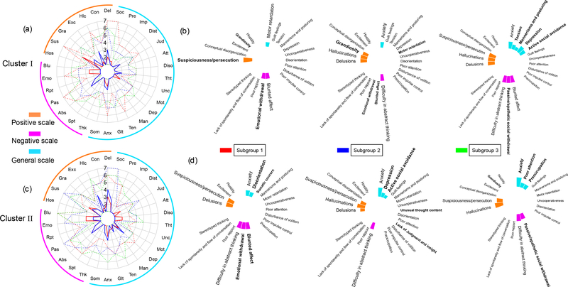
The extraction of common and distinct biomedical signatures among different populations allows for a more detailed study of the group-specific as well as distinct information of different populations. A number of subspace analysis algorithms have been developed and successfully applied to data fusion, however they are limited to joint analysis of only a couple of datasets. Since subspace analysis is very promising for analysis of multi-subject medical imaging data as well, we focus on this problem and propose a new method based on independent vector analysis (IVA) for common subspace extraction (IVA-CS) for multi-subject data analysis. IVA-CS leverages the strength of IVA in identification of a complete subspace structure across multiple datasets along with an efficient solution that uses only second-order statistics. We propose a subset analysis approach within IVA-CS to mitigate issues in estimation in IVA due to high dimensionality, both in terms of components estimated and the number of datasets. We introduce a scheme to determine a desirable size for the subset that is high enough to exploit the dependence across datasets and is not affected by the high dimensionality issue. We demonstrate the success of IVA-CS in extracting complex subset structures and apply the method to analysis of functional magnetic resonance imaging data from 179 subjects and show that it successfully identifies shared and complementary brain patterns from patients with schizophrenia (SZ) and healthy controls group. Two components with linked resting-state networks are identified to be unique to the SZ group providing evidence of functional dysconnectivity. IVA-CS also identifies subgroups of SZs that show significant differences in terms of their brain networks and clinical symptoms.
Keywords: Functional magnetic resonance imaging; Heterogeneity of schizophrenia; Independent vector analysis; Multi-subject medical imaging data; Subspace analysis.
Copyright © 2020 The Author(s). Published by Elsevier Inc. All rights reserved.
Figures
Similar articles
-
Constrained Independent Vector Analysis With Reference for Multi-Subject fMRI Analysis.IEEE Trans Biomed Eng. 2024 Dec;71(12):3531-3542. doi: 10.1109/TBME.2024.3432273. Epub 2024 Nov 21. IEEE Trans Biomed Eng. 2024. PMID: 39042541
-
A Scalable Approach to Independent Vector Analysis by Shared Subspace Separation for Multi-Subject fMRI Analysis.Sensors (Basel). 2023 Jun 5;23(11):5333. doi: 10.3390/s23115333. Sensors (Basel). 2023. PMID: 37300060 Free PMC article.
-
Task modulations and clinical manifestations in the brain functional connectome in 1615 fMRI datasets.Neuroimage. 2017 Feb 15;147:243-252. doi: 10.1016/j.neuroimage.2016.11.073. Epub 2016 Dec 1. Neuroimage. 2017. PMID: 27916665
-
Dynamic Reorganization of Functional Connectivity Reveals Abnormal Temporal Efficiency in Schizophrenia.Schizophr Bull. 2019 Apr 25;45(3):659-669. doi: 10.1093/schbul/sby077. Schizophr Bull. 2019. PMID: 29878254 Free PMC article.
-
Spatial Variance in Resting fMRI Networks of Schizophrenia Patients: An Independent Vector Analysis.Schizophr Bull. 2016 Jan;42(1):152-60. doi: 10.1093/schbul/sbv085. Epub 2015 Jun 23. Schizophr Bull. 2016. PMID: 26106217 Free PMC article.
Cited by
-
Constrained Independent Vector Analysis With Reference for Multi-Subject fMRI Analysis.IEEE Trans Biomed Eng. 2024 Dec;71(12):3531-3542. doi: 10.1109/TBME.2024.3432273. Epub 2024 Nov 21. IEEE Trans Biomed Eng. 2024. PMID: 39042541
-
Reproducibility in Matrix and Tensor Decompositions: Focus on Model Match, Interpretability, and Uniqueness.IEEE Signal Process Mag. 2022 Jul;39(4):8-24. doi: 10.1109/msp.2022.3163870. Epub 2022 Jun 28. IEEE Signal Process Mag. 2022. PMID: 36337436 Free PMC article. No abstract available.
-
Identification of Homogeneous Subgroups from Resting-State fMRI Data.Sensors (Basel). 2023 Mar 20;23(6):3264. doi: 10.3390/s23063264. Sensors (Basel). 2023. PMID: 36991975 Free PMC article.
-
Exploring synergies: Advancing neuroscience with machine learning.Signal Processing. 2026 Jan;238:110116. doi: 10.1016/j.sigpro.2025.110116. Epub 2025 Jun 2. Signal Processing. 2026. PMID: 40843337
-
A Scalable Approach to Independent Vector Analysis by Shared Subspace Separation for Multi-Subject fMRI Analysis.Sensors (Basel). 2023 Jun 5;23(11):5333. doi: 10.3390/s23115333. Sensors (Basel). 2023. PMID: 37300060 Free PMC article.
References
-
- Abolghasemi V, Ferdowsi S, Sanei S, 2015. Fast and incoherent dictionary learning algorithms with application to fMRI. Signal, Image and Video Processing 9 (1), 147–158.
-
- Adalı T, Anderson M, Fu G-S, May 2014. Diversity in independent component and vector analyses: Identifiability, algorithms, and applications in medical imaging. IEEE Signal Processing Magazine 31 (3), 18–33.
Publication types
MeSH terms
Grants and funding
LinkOut - more resources
Full Text Sources
Medical
