. 2020 Apr 28;10(5):684.
doi: 10.3390/biom10050684.

Culture and Differentiation of Human Hair Follicle Dermal Papilla Cells in a Soft 3D Self-Assembling Peptide Scaffold

Affiliations

Culture and Differentiation of Human Hair Follicle Dermal Papilla Cells in a Soft 3D Self-Assembling Peptide Scaffold

Nausika Betriu et al. Biomolecules. .

Abstract

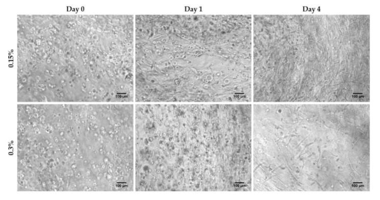
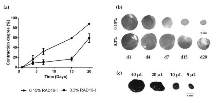
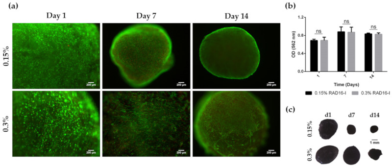
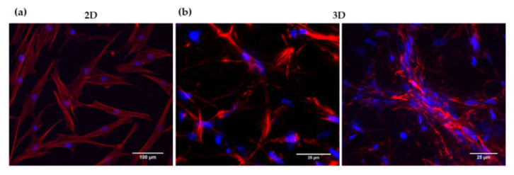
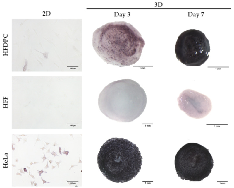
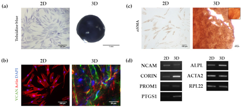
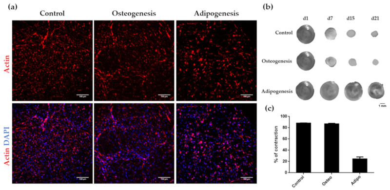
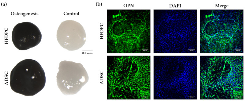
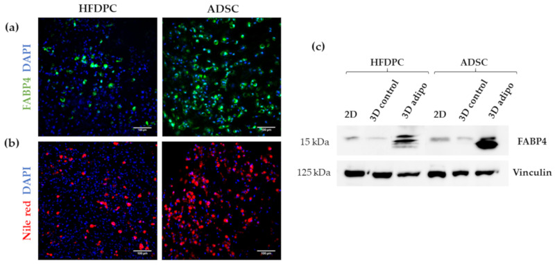
Hair follicle dermal papilla cells (HFDPC) are a specialized cell population located in the bulge of the hair follicle with unique characteristics such as aggregative behavior and the ability to induce new hair follicle formation. However, when expanded in conventional 2D monolayer culture, their hair inductive potency is rapidly lost. Different 3D culture techniques, including cell spheroid formation, have been described to restore, at least partially, their original phenotype, and therefore, their hair inductive ability once transplanted into a recipient skin. Moreover, hair follicle dermal papilla cells have been shown to differentiate into all mesenchymal lineages, but their differentiation potential has only been tested in 2D cultures. In the present work, we have cultured HFDPC in the 3D self-assembling peptide scaffold RAD16-I to test two different tissue engineering scenarios: restoration of HFDPC original phenotype after cell expansion and osteogenic and adipogenic differentiation. Experimental results showed that the 3D environment provided by RAD16-I allowed the restoration of HFDPC signature markers such as alkaline phosphatase, versican and corin. Moreover, RAD16-I supported, in the presence of chemical inductors, three-dimensional osteogenic and adipogenic differentiation. Altogether, this study suggests a potential 3D culture platform based on RAD16-I suitable for the culture, original phenotype recovery and differentiation of HFDPC.

Keywords: adipogenesis; hair follicle dermal papilla cells; osteogenesis; self-assembling peptides; tissue engineering.


Conflict of interest statement

The authors declare no conflict of interest.

Figures

Figure 1
Figure 1
HFDPC cultured in 3D self-assembling peptide scaffold RAD16-I at 0.15% and 0.3% peptide concentration. Scale bar 100 µm.
Figure 2
Figure 2
Matrix contraction of HFDPC in RAD16-I scaffolds. (a) Matrix contraction degree in soft (0.15%) and stiff (0.3%) hydrogels; (b) Macroscopic view of 0.15% and 0.3% peptide concentration 3D constructs along culture time; (c) 0.15% RAD16-I constructs at day 7 of culture stained by MTT. Construct size can be modulated by changing the initial seeding volume.
Figure 3
Figure 3
Viability of HFDPC cultured in the 3D self-assembling peptide scaffold RAD16-I at 0.15% and 0.3% peptide concentration. (a) Fluorescent images of Live/Dead staining. Scale bar: 200 µm; (b) MTT absorbance values of 3D HFDPC constructs at different time points in culture; (c) Macroscopic view of 3D constructs after MTT incubation.
Figure 4
Figure 4
DAPI/Phalloidin staining of HFDPC in (a) 2D and (b) 3D cultures.
Figure 5
Figure 5
Alkaline phosphatase staining in 2D and 3D cultures. HFDPC were negatively stained for ALP activity in 2D cultures. The expression of ALP was recovered once encapsulated in RAD16-I 3D scaffolds. Human Foreskin Fibroblasts (HFF) were used as a negative control and HeLa cells as a positive control for ALP staining.
Figure 6
Figure 6
Analysis of HFDPC markers in 2D and RAD16-I 3D cultures. (a) Toluidine blue staining for the detection of glycosaminoglycans; (b) Versican (VCAN) immunofluorescence. VCAN was absent in 2D cultures but its expression was recovered in 3D cultures as shown by immunofluorescence detection; (c) α Smooth Muscle Actin (αSMA) immunocytochemistry. αSMA was detected in both 2D and 3D cultures. The inner box represents a negative control (incubation with secondary antibody only); (d) Agarose gel of Reverse-transcription PCR products including Neural Cell Adhesion Molecule (NCAM), Corin (CORIN), Prominin-1 (PROM1), Prostaglandin endoperoxidase synthase 1 (PTGS1), alkaline phosphatase (ALPL) and α-Smooth Muscle Actin (ACTA2).
Figure 7
Figure 7
Influence of osteogenic and adipogenic induction on cell morphology and matrix contraction in HFDPC. (a) DAPI/Phalloidin staining of 3D cultures under control, osteogenic and adipogenic medium; (b) Construct contraction along culture time; (c) Contraction degree after 21 days of culture under control, osteogenic and adipogenic medium.
Figure 8
Figure 8
Characterization of osteogenic phenotype of HFDPC and ADSC cultured in RAD16-I scaffold for 21 days. (a) von Kossa staining (calcium mineralization) of 3D constructs cultured in control and osteogenic medium; (b) OPN immunofluorescence of 3D constructs under osteogenic induction.
Figure 9
Figure 9
Characterization of adipogenic phenotype of HFDPC and ADSC cultured in RAD16-I scaffold for 21 days. (a) FABP4 immunofluorescence of 3D constructs under adipogenic induction; (b) Nile red staining of 3D constructs under adipogenic induction; (c) FABP4 western blot of HFDPC and ADSC cultured in 2D cultures and in 3D RAD16-I scaffold under control and adipogenic medium. Vinculin was used as an internal control. The image is representative of three replicates.

References

    1. Teumer J., Cooley J. Follicular cell implantation: An emerging cell therapy for hair loss. Semin. Plast. Surg. 2005;19:193–200. doi: 10.1055/s-2005-871735. - DOI
    1. Driskell R.R., Clavel C., Rendl M., Watt F.M. Hair follicle dermal papilla cells at a glance. J. Cell Sci. 2011;124:1179–1182. doi: 10.1242/jcs.082446. - DOI - PMC - PubMed
    1. Yang C.C., Cotsarelis G. Review of hair follicle dermal cells. J. Dermatol. Sci. 2010;57:2–11. doi: 10.1016/j.jdermsci.2009.11.005. - DOI - PMC - PubMed
    1. Jahoda C.A.B., Reynolds A.J., Chaponnier C., Forester J.C., Gabbiani G. Smooth muscle a-actin is a marker for hair follicle dermis In Vivo and In Vitro. J. Cell Sci. 1991;99:627–636. - PubMed
    1. Qiao J., Zawadzka A., Philips E., Turetsky A., Batchelor S., Peacock J., Durrant S., Garlick D., Kemp P., Teumer J. Hair follicle neogenesis induced by cultured human scalp dermal papilla cells. Regen. Med. 2009;4:667–676. doi: 10.2217/rme.09.50. - DOI - PubMed

Publication types

LinkOut - more resources