. 2020 Aug 4;142(5):483-498.
doi: 10.1161/CIRCULATIONAHA.119.044803. Epub 2020 May 1.

Cyclodextrin Prevents Abdominal Aortic Aneurysm via Activation of Vascular Smooth Muscle Cell Transcription Factor EB

Affiliations

Cyclodextrin Prevents Abdominal Aortic Aneurysm via Activation of Vascular Smooth Muscle Cell Transcription Factor EB

Haocheng Lu et al. Circulation. .

Erratum in

Abstract

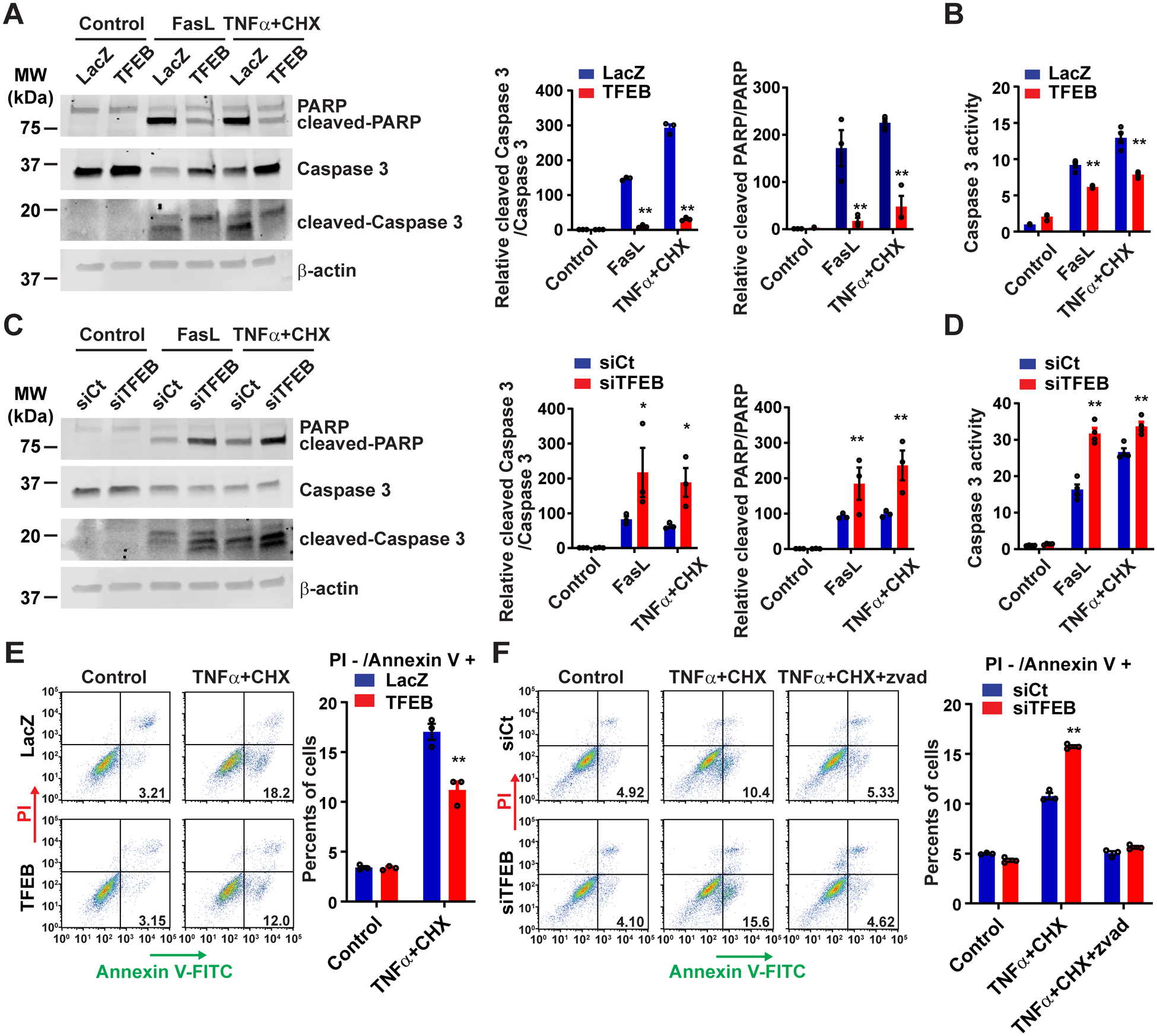
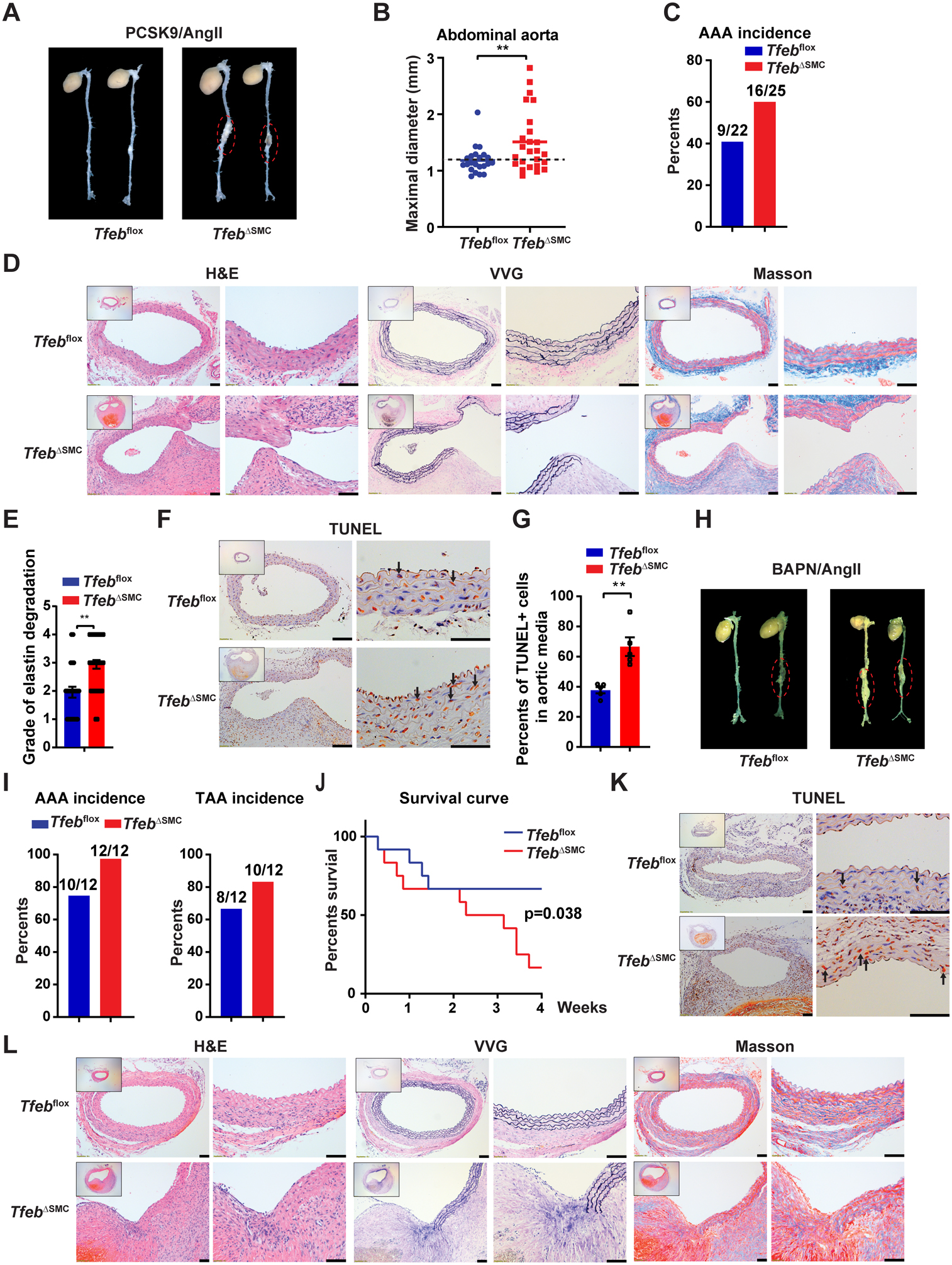
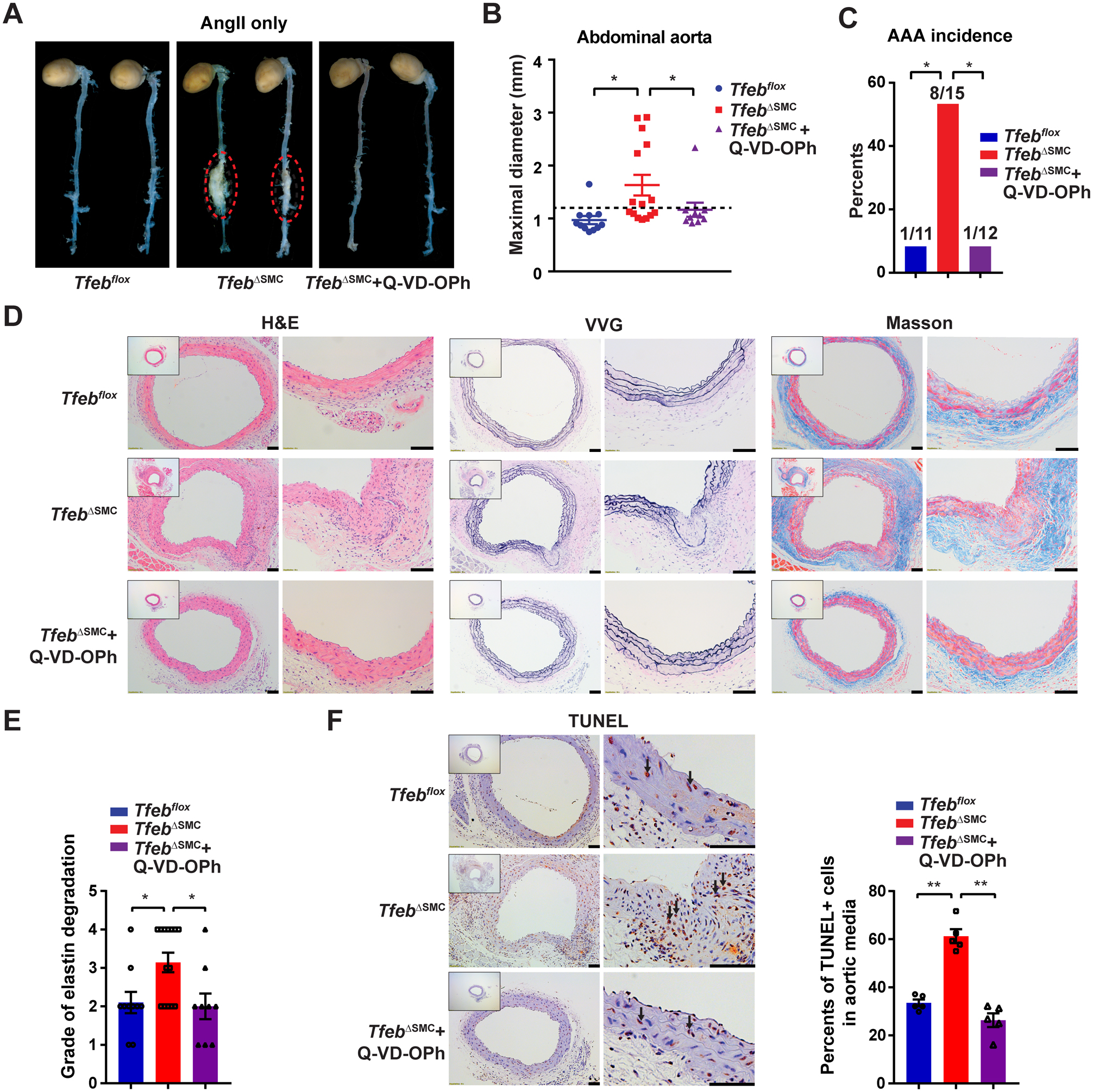
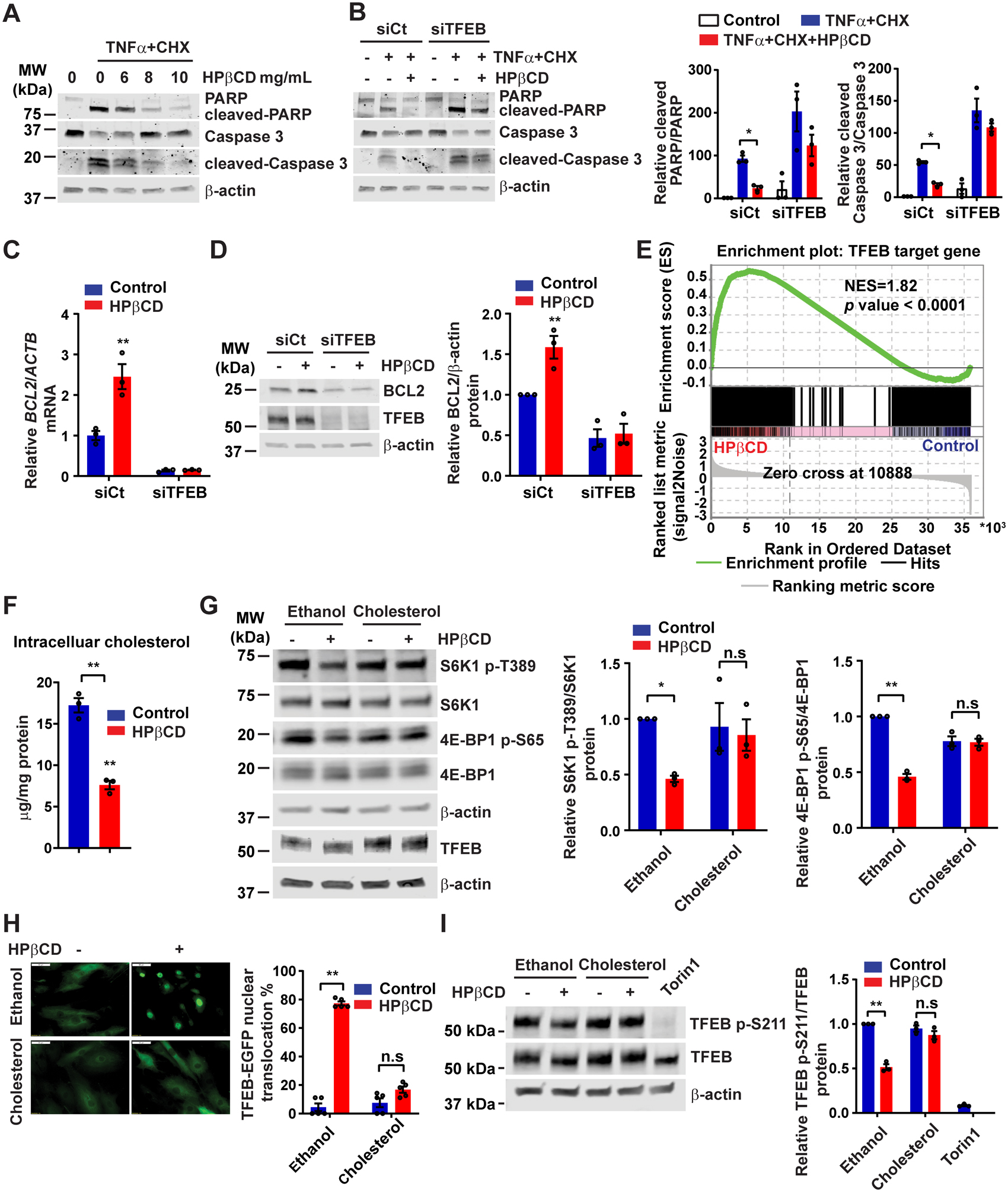
Background: Abdominal aortic aneurysm (AAA) is a severe aortic disease with a high mortality rate in the event of rupture. Pharmacological therapy is needed to inhibit AAA expansion and prevent aneurysm rupture. Transcription factor EB (TFEB), a master regulator of autophagy and lysosome biogenesis, is critical to maintain cell homeostasis. In this study, we aim to investigate the role of vascular smooth muscle cell (VSMC) TFEB in the development of AAA and establish TFEB as a novel target to treat AAA.

Methods: The expression of TFEB was measured in human and mouse aortic aneurysm samples. We used loss/gain-of-function approaches to understand the role of TFEB in VSMC survival and explored the underlying mechanisms through transcriptome and functional studies. Using VSMC-selective Tfeb knockout mice and different mouse AAA models, we determined the role of VSMC TFEB and a TFEB activator in AAA in vivo.

Results: We found that TFEB is downregulated in both human and mouse aortic aneurysm lesions. TFEB potently inhibits apoptosis in VSMCs, and transcriptome analysis revealed that TFEB regulates apoptotic signaling pathways, especially apoptosis inhibitor B-cell lymphoma 2. B-cell lymphoma 2 is significantly upregulated by TFEB and is required for TFEB to inhibit VSMC apoptosis. We consistently observed that TFEB deficiency increases VSMC apoptosis and promotes AAA formation in different mouse AAA models. Furthermore, we demonstrated that 2-hydroxypropyl-β-cyclodextrin, a clinical agent used to enhance the solubility of drugs, activates TFEB and inhibits AAA formation and progression in mice. Last, we found that 2-hydroxypropyl-β-cyclodextrin inhibits AAA in a VSMC TFEB-dependent manner in mouse models.

Conclusions: Our study demonstrated that TFEB protects against VSMC apoptosis and AAA. TFEB activation by 2-hydroxypropyl-β-cyclodextrin may be a promising therapeutic strategy for the prevention and treatment of AAA.

Keywords: aortic aneurysm, abdominal; apoptosis; autophagy; myocytes, smooth muscle.


Conflict of interest statement

Disclosures: Authors declare no competing interests.

Figures

Figure 1.
Figure 1.. TFEB is downregulated in human and mouse aortic aneurysm.
(A) Transcription factor EB (TFEB) mRNA and (B) protein were determined by quantitative polymerase chain reaction (qPCR) and Western blot, respectively, in human proximal ascending aortic aneurysm lesions compared with that in the adjacent normal aorta (n=12 for mRNA, n=8 for protein). (C) TFEB was detected by immunofluorescence in human aneurysmal lesions and adjacent normal aorta. Scale bar, 50 μm. (D) Human aortic smooth muscle cells (HASMCs) were treated with tumor necrosis factor α (TNFα; 20 ng/mL), interleukin 1 β (IL1β; 10 ng/mL) or Interferon γ (IFNγ; 50 ng/mL) for 72 hours. TFEB protein was determined by Western blot (n=3). (E-F) In the proprotein convertase subtilisin/kexin type 9 (PCSK9)/ angiotensin II (AngII) model, TFEB mRNA and protein were determined in the aortas of AngII- and saline-infused mice by qPCR (E; n=15 for saline, n=8 for AngII) or Western blot (F; n=6). Data are presented as mean ± SEM. *p < 0.05, **p < 0.01. Paired t-test for A and B, one-way ANOVA followed by Holm-Sidak post hoc analysis for D, and unpaired t-test for E and F.
Figure 2.
Figure 2.. TFEB inhibits apoptosis in human aortic smooth muscle cells (HASMCs).
(A-B) HASMCs were infected with adenovirus encoding LacZ (Ad-LacZ) or TFEB (Ad-TFEB) (multiplicity of infection [MOI], 30). After 48 hours, the cells were treated with Fas ligand (FasL; 100 ng/mL) or tumor necrosis factor α (TNFα; 100 ng/mL) + cycloheximide (CHX; 20 μg/mL) for 6 hours. The apoptosis was assessed by Western blot (A) and cysteine-aspartic protease, cysteine aspartase (Caspase 3) activity assay (B). (C-D) HASMCs were transfected with siRNA-negative control (siCt; 30 nM) or siRNA-TFEB (siTFEB; 30 nM). After 72 hours, the cells were treated with FasL or TNFα+CHX for 6 hours. Similarly, the apoptosis was determined by Western blot (C) or Caspase 3 activity assay (D). (E-F) HASMCs were infected with Ad-LacZ or Ad-TFEB (MOI, 30) for 48 hours (E) or transfected with siCt or siTFEB (30 nM) for 72 hours (F), and then the cells were treated with TNFα + CHX for 4 hours before Annexin V/propidium iodide (PI) staining and flow cytometry analysis. Data are presented as mean ± SEM of three independent experiments. *p < 0.05, **p < 0.01 compared with Ad-LacZ or siCt group. Two-way ANOVA followed by Holm-Sidak post hoc analysis for A, B, C, D, E, F. Zvad: carbobenzoxy-valyl-alanyl-aspartyl-[O-methyl]- fluoromethylketone
Figure 3.
Figure 3.. TFEB inhibits apoptosis through upregulation of B-cell lymphoma 2 (BCL2) in human aortic smooth muscle cells (HASMCs).
(A and B) HASMCs were infected with adenovirus encoding LacZ (Ad-LacZ) or Ad-TFEB (MOI, 30) for 48 hours or (C and D) HASMCs were transfected with siRNA-control (siCt; 30 nM) or siRNA-TFEB (siTFEB; 30 nM) for 72 hours, BCL2 mRNA and protein were determined by quantitative polymerase chain reaction (qPCR) (A, C) or Western blot (B, D). (E) Chromatin immunoprecipitation (ChIP) assay was performed in HASMCs infected with Ad-LacZ or Ad-flag-TFEB (MOI, 30). The binding of TFEB to the BCL2 promoter was determined by qPCR. (F) CV1 cells were transfected with a human BCL2 promoter-driven luciferase vector containing a wild type (WT) or mutated TFEB binding site. After 24 hours, the cells were infected with Ad-LacZ and Ad-TFEB (MOI, 30). The luciferase activity was measured and normalized to Renilla activity. (G) HASMCs were infected with Ad-LacZ and Ad-TFEB (MOI, 30). After 48 hours, the cells were pretreated with ABT199 (200 nM) or HA14–1 (20 μM) for 1 hour followed by treatment with TNFα + CHX for 6 hours. Apoptosis was determined by Western blot. Data are presented as mean ± SEM of three independent experiments. *p < 0.05, **p < 0.01 compared with Ad-LacZ, siCt or IgG group. Unpaired t-test for A, B, C, D and two-way ANOVA followed by Holm-Sidak post hoc analysis for E, F and G.
Figure 4.
Figure 4.. Vascular smooth muscle cell (VSMC)-Tfeb KO aggravates abdominal aortic aneurysm (AAA) formation in the proprotein convertase subtilisin/kexin type 9 (PCSK9)/ angiotensin II (AngII) and β-aminopropionitrile (BAPN)/AngII models.
(A-G) In the PCSK9/AngII model, Tfebflox and TfebΔSMC mice were injected (i.p.) with adeno-associated virus (AAV)-PCSK9 D337Y and fed a western diet. After 2 weeks, the mice were infused with AngII (1,000 ng/kg/min) for another 4 weeks. (A) Representative images of AAA. (B) Maximal diameter of the abdominal aorta. (C) AAA incidence. (D) Representative hematoxylin and eosin (H&E), Verhoeff-Van Gieson (VVG) and Masson’s trichrome staining of the mouse aorta. (E) Grade of elastin degradation in the aortic wall. (F-G) Terminal deoxynucleotidyl transferase dUTP nick end labeling (TUNEL) staining of the aortic wall. Normal cell nuclei are blue; apoptotic cell nuclei pare brown. Arrows point to apoptotic cells. The percentage of TUNEL positive nuclei in the aortic media was determined. In (A-E), n=22 for Tfebflox mice and n=25 for TfebΔSMC mice. (H-L) In the BAPN/AngII model, Tfebflox and TfebΔSMC mice were infused with both AngII (1,000 ng/kg/min, 4 weeks) and BAPN (150 mg/kg/min, during the first 2 weeks). (H) Representative images of AAA. (I) AAA and TAA incidence. (J) Survival curve. (K) TUNEL staining of the aortic wall. (L) Representative H&E, VVG and Masson’s trichrome staining of the mouse aorta. In (H-L), n=12 for each genotype. Low-magnification images in D, F, K and L show the entire vascular wall at the site of analysis. Data are presented as mean ± SEM. **p < 0.01. Mann-Whitney test for B, unpaired t-test for E and G, and Mantel-Cox method for J. Scale bar, 50 μm.
Figure 5.
Figure 5.. Vascular smooth muscle cell (VSMC)-Tfeb KO promotes abdominal aortic aneurysm (AAA) in angiotensin II (AngII) infusion-induced hypertension model.
WT (Tfebflox) mice and VSMC-Tfeb KO (TfebΔSMC) mice were infused with AngII (1,000 ng/kg/min) subcutaneously for 8 weeks to induce AAA, and simultaneously administered vehicle control (DMSO) or apoptosis inhibitor Q-VD-OPh (20 mg/kg daily, i.p.) starting one day before AngII infusion. (A) Representative images of the mouse AAA. (B) Maximal diameter of the abdominal aorta. (C) AAA incidence. (D) Representative H&E, VVG and Masson’s trichrome staining of the mouse aorta. (E) Grade of the elastin degradation in the aortic wall. (F) The apoptotic cells were assessed by terminal deoxynucleotidyl transferase dUTP nick end labeling (TUNEL) staining in the aortic wall. Normal cell nuclei are blue; apoptotic cell nuclei are brown. Arrows point to apoptotic cells. The percentage of TUNEL positive nuclei in the aortic media was determined. Low-magnification images in D and F show the entire vascular wall at the site of analysis. Data are presented as mean ± SEM. *p < 0.05, **p < 0.01. Kruskal-Wallis test followed by a two-stage step-up method of Benjamini, Krieger and Yekutieli for B, Fisher’s exact test for C, one-way ANOVA followed by Holm-Sidak post hoc analysis for E and F. (A-E) n=11, 15, 12 for the Tfebflox, TfebΔSMC and TfebΔSMC + Q-VD-OPh group, respectively. Scale bar, 50 μm.
Figure 6.
Figure 6.. 2-hydroxypropyl-beta-cyclodextrin (HPβCD) activates TFEB by reducing intracellular cholesterol.
(A) Human aortic smooth muscle cells (HASMCs) were treated with HPβCD at the indicated concentration for 24 hours and then treated with tumor necrosis factor α (TNFα; 100 ng/mL) + cycloheximide (CHX; 20 μg/mL) for 6 hours. The cleavage of poly ADP-ribose polymerase (PARP) and Caspase 3 was determined by Western blot. (B) HASMCs were transfected with siCt or siTFEB (30 nM). After 48 hours, the cells were treated with HPβCD (10 mg/mL) for 24 hours and subsequently treated with TNFα + CHX as in (A). (C-D) HASMCs were transfected with siCt or siTFEB (30 nM). After 48 hours, the cells were treated with HPβCD (10 mg/mL) for 24 hours. B-cell lymphoma 2 (BCL2) expression was determined by quantitative polymerase chain reaction (qPCR) (C) and Western blot (D). (E) HASMCs were treated with HPβCD (10 mg/mL) for 24 hours and RNA was extracted for RNA-sequencing. Gene Set Enrichment Analysis (GSEA) was performed to determine the enrichment of TFEB target genes in the HPβCD-treated group. (F) Intracellular cholesterol was measured in the HASMCs treated with HPβCD (10 mg/mL) for 24 hours. (G) HASMCs were treated with HPβCD (10 mg/mL) in the presence or absence of cholesterol (20 μg/mL) for 6 hours. Mechanistic target of rapamycin complex 1 (mTORC1) activity was determined by Western blot. (H) HASMCs were infected with adenovirus encoding TFEB-enhanced green fluorescent protein (Ad-TFEB-EGFP; MOI, 20), and 24 hours later, the cells were treated with HPβCD (10 mg/mL) in the presence or absence of cholesterol (20 μg/mL) for 6 hours. The TFEB nuclear translocation was analyzed by fluorescent microscopy. (I) HASMCs were infected with Ad-TFEB (MOI, 10), and 48 hours later, the cells were treated with HPβCD (10 mg/mL) in the presence or absence of cholesterol (20 μg/mL) for 6 hours. The phosphorylation of TFEB on Ser211 was determined by Western blot. Data are presented as mean ± SEM of three independent experiments. *p < 0.05, **p < 0.01; n.s: not significant. Two-way ANOVA followed by Holm-Sidak post hoc analysis for B, C, D, G, H, I and unpaired t-test for F. Scale bar, 50 μm.
Figure 7.
Figure 7.. 2-hydroxypropyl-beta-cyclodextrin (HPβCD) inhibits abdominal aortic aneurysm (AAA) formation in a vascular smooth muscle cell (VSMC) TFEB-dependent manner.
(A-E) In the proprotein convertase subtilisin/kexin type 9 (PCSK9)/angiotensin II (AngII) model, after inducing hyperlipidemia by AAV-PCSK9 D337Y and western diet, C57BL/6J mice were infused with AngII (1,500 ng/kg/min) for another 4 weeks. Saline (vehicle control) or HPβCD (2 g/kg) were administered (i.p.) twice a week, starting one day before AngII infusion. (A) Representative images of abdominal aortic aneurysm (AAA). (B) Maximal diameter of the abdominal aorta. (C) AAA incidence. (D) Representative H&E, VVG and Masson’s trichrome staining of the mouse aortas. (E) Terminal deoxynucleotidyl transferase dUTP nick end labeling (TUNEL) staining of the aortic wall. In (A-D) n=10 for each group. Low-magnification images in D and E show the entire vascular wall of normal aorta or AAA. (F) To study the aortic aneurysm progression, AAA was first induced in C57BL/6J mice by the method of PCSK9/AngII (1,500 ng/kg/min). The mice were then randomly divided into 2 groups: saline and HPβCD (2 g/kg, i.p. twice a week). The aorta was assessed by ultrasound at week 4 and week 8 after AngII infusion. The change in the internal diameter of the suprarenal aorta was calculated as [diameter (week 8)] – [diameter (week 4)]. In (F), n=9 for each group. (G-J) In PCSK9/AngII model, Tfebflox mice and TfebΔSMC were treated with either saline or HPβCD (2 g/kg, i.p.) starting one day before AngII infusion. (G) Representative images of AAA. (H) Maximal diameter of the abdominal aorta. (I) AAA incidence. (J) TUNEL staining of the aortic wall. In (G-I), n=19 for Tfebflox + Saline, n=12 for Tfebflox + HPβCD, n=16 for TfebΔSMC + Saline and n=13 for TfebΔSMC + HPβCD. (K) Proposed model for TFEB as a therapeutic target in AAA. Data are presented as mean ± SEM. *p < 0.05, **p < 0.01; n.s, not significant. Mann-Whitney test for B, unpaired t-test for E and F, Kruskal-Wallis test followed by two-stage step-up method of Benjamini, Krieger and Yekutieli for H, and two-way ANOVA followed by Holm-Sidak post hoc analysis for J. Scale bar, 50 μm.

References

    1. Kent KC. Clinical practice. Abdominal aortic aneurysms. New Eng J Med. 2014;371:2101–2108. doi: 10.1056/NEJMcp1401430 - DOI - PubMed
    1. Chaikof EL, Dalman RL, Eskandari MK, Jackson BM, Lee WA, Mansour MA, Mastracci TM, Mell M, Murad MH, Nguyen LL, et al. The Society for Vascular Surgery practice guidelines on the care of patients with an abdominal aortic aneurysm. J Vasc Surg. 2018;67:2–77 e2. doi: 10.1016/j.jvs.2017.10.044 - DOI - PubMed
    1. Rowe VL, Stevens SL, Reddick TT, Freeman MB, Donnell R, Carroll RC and Goldman MH. Vascular smooth muscle cell apoptosis in aneurysmal, occlusive, and normal human aortas. J Vasc Surg. 2000;31:567–576. doi: 10.1067/mva.2000.102847 - DOI - PubMed
    1. Settembre C, Di Malta C, Polito VA, Garcia Arencibia M, Vetrini F, Erdin S, Erdin SU, Huynh T, Medina D, Colella P, et al. TFEB links autophagy to lysosomal biogenesis. Science. 2011;332:1429–1433. doi: 10.1126/science.1204592 - DOI - PMC - PubMed
    1. Decressac M and Bjorklund A. TFEB: Pathogenic role and therapeutic target in Parkinson disease. Autophagy. 2013;9:1244–1246. doi: 10.4161/auto.25044 - DOI - PMC - PubMed

Publication types

MeSH terms