. 2020 Jul 31;127(4):453-465.
doi: 10.1161/CIRCRESAHA.119.316448. Epub 2020 May 1.

Gut Microbiota-Derived Short-Chain Fatty Acids Promote Poststroke Recovery in Aged Mice

Affiliations

Gut Microbiota-Derived Short-Chain Fatty Acids Promote Poststroke Recovery in Aged Mice

Juneyoung Lee et al. Circ Res. .

Abstract

Rationale: The elderly experience profound systemic responses after stroke, which contribute to higher mortality and more severe long-term disability. Recent studies have revealed that stroke outcomes can be influenced by the composition of gut microbiome. However, the potential benefits of manipulating the gut microbiome after injury is unknown.

Objective: To determine if restoring youthful gut microbiota after stroke aids in recovery in aged subjects, we altered the gut microbiome through young fecal transplant gavage in aged mice after experimental stroke. Further, the effect of direct enrichment of selective bacteria producing short-chain fatty acids (SCFAs) was tested as a more targeted and refined microbiome therapy.

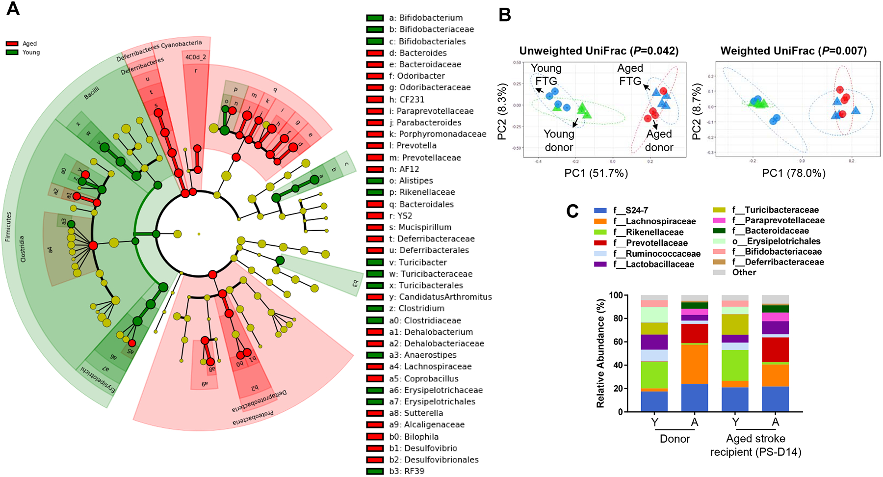
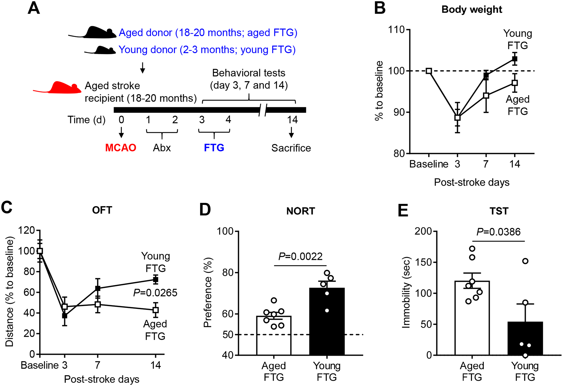
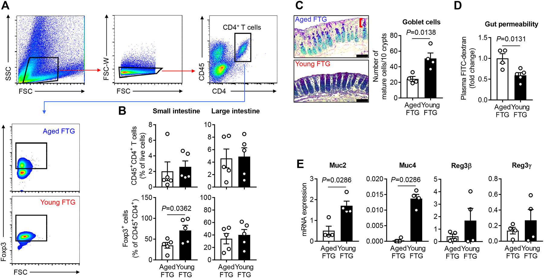
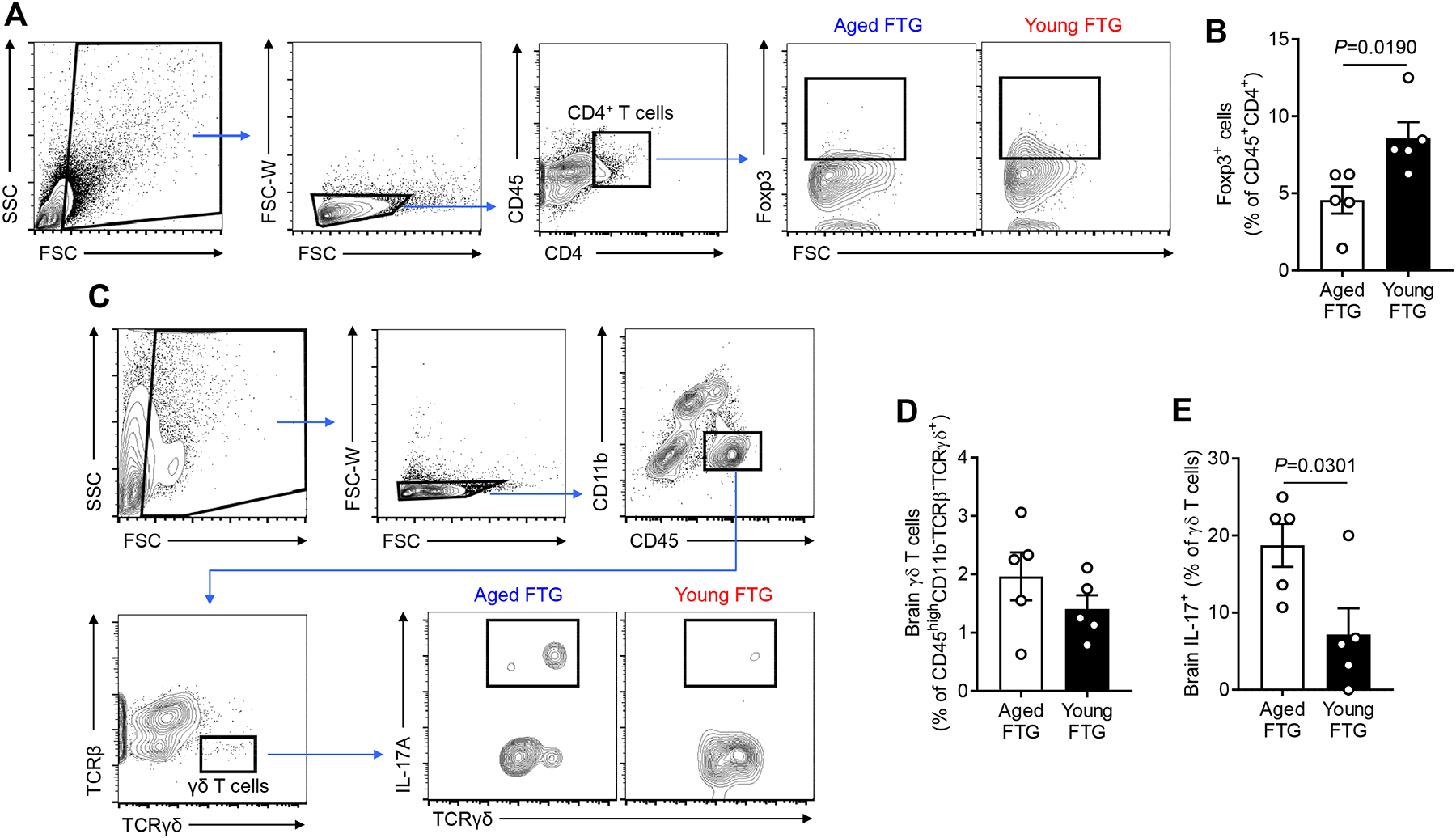
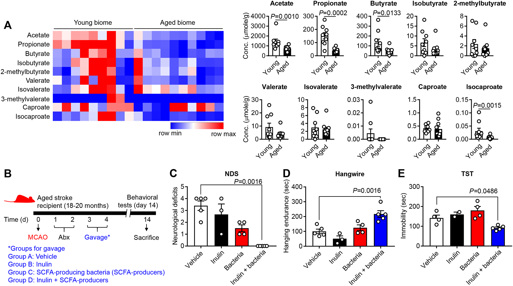
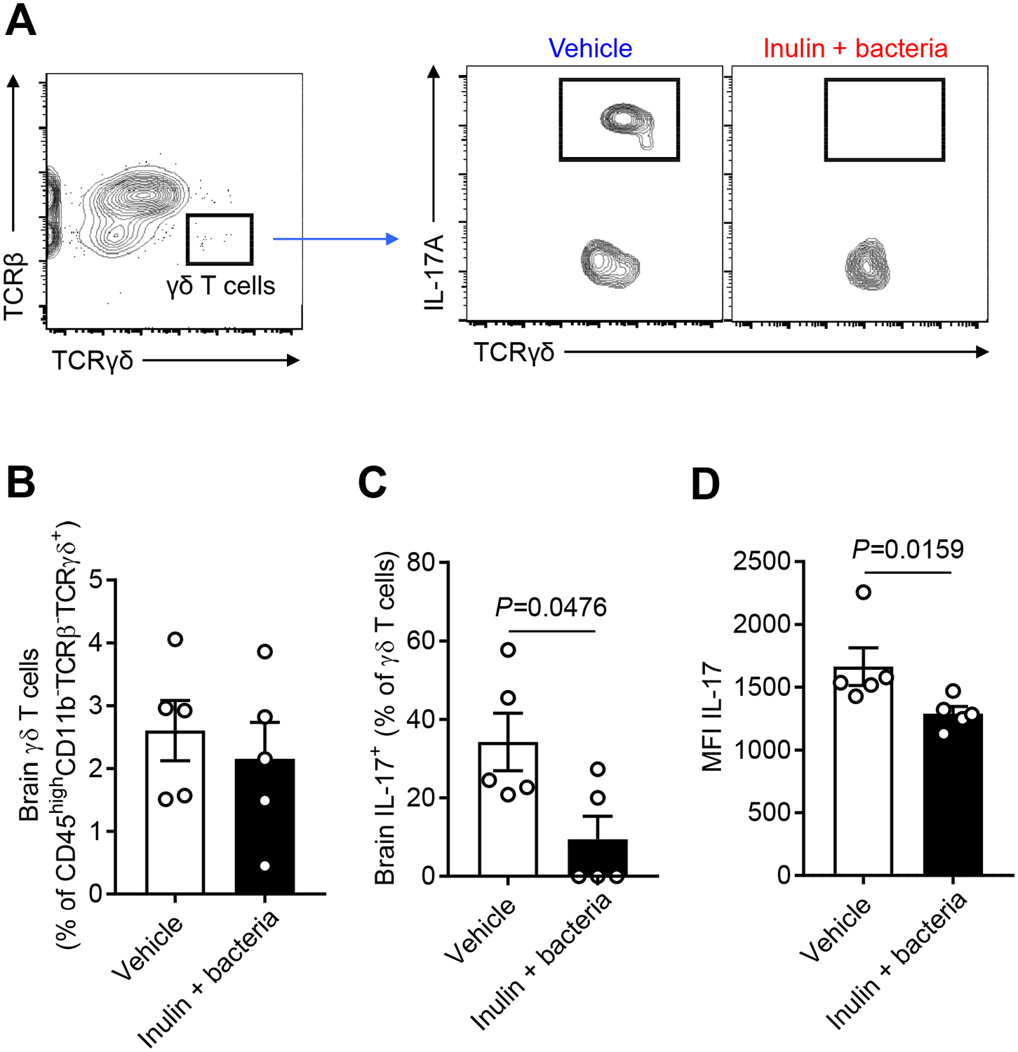
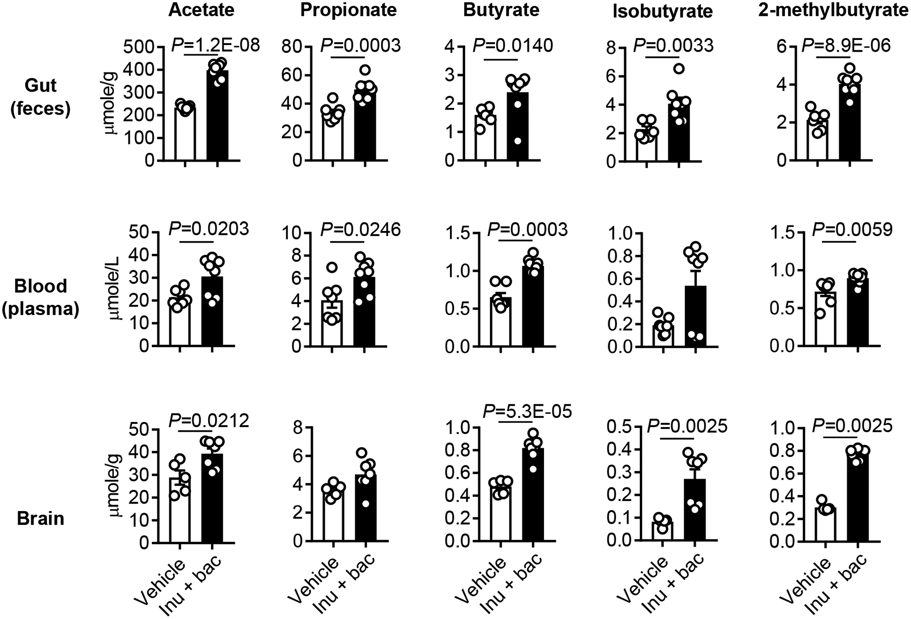
Methods and results: Aged male mice (18-20 months) were subjected to ischemic stroke by middle cerebral artery occlusion. We performed fecal transplant gavage 3 days after middle cerebral artery occlusion using young donor biome (2-3 months) or aged biome (18-20 months). At day 14 after stroke, aged stroke mice receiving young fecal transplant gavage had less behavioral impairment, and reduced brain and gut inflammation. Based on data from microbial sequencing and metabolomics analysis demonstrating that young fecal transplants contained much higher SCFA levels and related bacterial strains, we selected 4 SCFA-producers (Bifidobacterium longum, Clostridium symbiosum, Faecalibacterium prausnitzii, and Lactobacillus fermentum) for transplantation. These SCFA-producers alleviated poststroke neurological deficits and inflammation, and elevated gut, brain and plasma SCFA concentrations in aged stroke mice.

Conclusions: This is the first study suggesting that the poor stroke recovery in aged mice can be reversed via poststroke bacteriotherapy following the replenishment of youthful gut microbiome via modulation of immunologic, microbial, and metabolomic profiles in the host.

Keywords: aging; fecal microbiota transplantation; gut microbiota; inflammation; metabolomics; middle cerebral artery occlusion.


Figures

Figure 1.
Figure 1.. Composition of the fecal microbiota of young and aged donor mice and clustering in recipient aged stroke mice.
A, Overall representation of bacterial profiles in young (2–3 months) and aged (18–20 months) donor mice at the baseline, by linear discriminant effect size (LEfSe) analysis (n=4 per group). B, Principal coordinates analysis (PCoA) of fecal microbiota from the donor (young: 2–3 months, aged: 18–20 months) and recipient stroke mice (18–20 months) with fecal transplant gavage (FTG) using unweighted (R2=0.2567) and weighted (R2=0.4562) UniFrac distances (n=4 per group). C, Bacterial composition at the family level in transplanted recipients cluster according to the donor profiles at day 14 post-MCAO (n=4 per group).
Figure 2.
Figure 2.. Post-stroke transplanting of young fecal microbiome improves behavior in aged mice.
A, The experimental protocol for FTG from young (2–3 months) and aged (18–20 months) donor mice into recipient aged stroke mice (18–20 months). Changes of body weight (B), total locomotor activity using open field test (OFT, C), cognitive function using novel objective recognition test (NORT, D) and depression-like behaviors using tail suspension test (TST, E) after young (n=5) and aged FTG (n=7) on day 14 following MCAO. Throughout, error bars represent mean±SEM. For the repeated measurement study, a linear mixed model was used to account for within-subject correlation (B and C). Group comparisons were further performed at each time adjusted for multiple testing. For two-group comparisons, Student’s t test was used after the normality of data was confirmed by the Shapiro-Wilk normality test (D and E).
Figure 3.
Figure 3.. Young FTG increases intestinal regulatory T (Treg) cells in the lamina propria (LP) and enhances mucin productions in the epithelium of aged stroke mice.
A, Representative flow cytometry plots to identify Treg cells in the intestinal LP of aged stroke mice. CD45+CD4+Foxp3+ cells in the LP of both small intestine (SI) and large intestine (LI) were gated and analyzed as Treg cells. An amine reactive Live/Dead Aqua viability stain was used to identify live and dead cells. Only live cells were gated for the analysis. Graphs represent percentages of Foxp3+ cells of CD4+ T cells in the SI LP. B, Flow cytometric analysis of CD4+ T cells and Treg cells in the intestinal LP of aged stroke mice at day 14 after MCAO (n=5 per group). C, Alcian blue and periodic acid-Schiff (AB-PAS) staining of the LI of aged stroke mice at post-MCAO day 14 (n=4 per group) and the number of mature goblet cells per 10 upper crypts/mouse are quantified. The bracket indicates the upper crypt possessing mature cells. Scale bars, 50 μm. D, FITC-dextran intestinal permeability assay at day 14 after MCAO in aged mice with aged (n=4) and young FTG (n=5). Data were normalized to aged FTG controls. E, The relative mRNA abundance for epithelial mucin genes and Reg3 genes of the LI in aged stroke mice at post-MCAO day 14 (n=4 per group). Throughout, error bars represent mean±SEM. Student’s t test (B, C and D) and Mann-Whitney U test (E) were used based on the normality of data assessed by the Shapiro-Wilk normality test.
Figure 4.
Figure 4.. Young FTG increases Treg cells and reduces IL-17 production of γδ T cells in the brain of aged stroke mice.
A, Representative flow cytometry plots to identify Treg cells (CD45+CD4+Foxp3+) in the brain of aged stroke mice. B, Flow cytometric analysis of Treg cells in the brain of aged stroke mice at day 14 after MCAO (n=5 per group). C, Representative flow cytometry plots to identify γδ T cells (CD45highCD11bTCRβTCRγδ+) in the brain of aged stroke mice. IL-17+ γδ T cells (CD45highCD11bTCRβTCRγδ+IL-17+) were analyzed at post-MCAO day 14. An amine reactive Live/Dead Aqua viability stain was used to identify live and dead cells. The ipsilateral stroke hemisphere was used for the cell isolation and only live cells were gated for the analysis for both (A) and (C). Flow cytometric analysis of brain γδ T cells (D) and IL-17+ γδ T cells (E) of aged stroke mice at day 14 after MCAO (n=5 per group). Throughout, error bars represent mean±SEM. Student’s t test (B, D and E) was used after the normality of data was confirmed by the Shapiro-Wilk normality test.
Figure 5.
Figure 5.. Fecal SCFA concentrations decrease with age and the combined treatment of SCFA-producing bacteria and prebiotic inulin after stroke ameliorates behavioral disorders in aged mice.
A, Aging decreases the concentration of primary SCFAs in fecal samples of young (2–3 months) and aged (18–20 months) mice. Fecal samples were collected and analyzed using mass spectrometry. Graphs represent changes of individual SCFAs (young biome, n=9; aged biome, n=10). B, To examine the effect of inulin and SCFA-producers on stroke outcome, four separate groups of mice were orally gavaged with 1) vehicle, 2) inulin, 3) SCFA-producers and 4) inulin+SCFA-producers. For SCFA-producers, the cocktail of Bifidobacterium longum (1×10⁷), Clostridium symbiosum (5×10⁶), Faecalibacterium prausnitzii (1×10⁶) and Lactobacillus fermentum (1×10⁹) were freshly cultured and gavaged to mice. Neurological deficit score (NDS) (C), hangwire test (D) and TST (E) were performed at day 14 after MCAO (vehicle, n=5; inulin alone, n=3; SCFA-producers alone, n=4; inulin+SCFA-producers, n=5). For the TST, two mice from vehicle- and inulin-treated group (one for each group) which showed spinning due to severe neurological deficits were excluded. Overall P value for NDS, hangwire test and TST was 0.0001, 0.0004 and 0.0036, respectively. Throughout, error bars represent mean±SEM. For group comparisons, Mann-Whitney U test (A) and ordinary one-way ANOVA or Kruskal-Wallis test (C, D and E) were used based on the normality of data assessed by the Shapiro-Wilk normality test, followed by Dunnett’s multiple comparison test.
Figure 6.
Figure 6.. Transplanting SCFA-producers reduces brain IL-17 production of γδ T cells in the aged stroke mice.
A, Representative flow cytometry plots to identify γδ T cells in the brain (ipsilateral stroke hemisphere) of aged stroke mice. The same gating strategy used in FTG study was employed. Brain γδ T cells (B) and IL-17+ γδ T cells (C) of aged stroke mice were analyzed at day 14 after MCAO (n=5 per group). The IL-17 median fluorescence intensity (MFI) was calculated (D). Throughout, error bars represent mean±SEM. Mann-Whitney U test (B, C and D) was used because the normality of data was rejected by the Shapiro-Wilk normality test.
Figure 7.
Figure 7.. SCFA profiles in SCFA-producers-treated aged stroke mice.
At day 14 after MCAO and Abx (days 1 and 2) and inulin+bacteria (days 3 and 4) treatment, the samples were collected from aged stroke mice. Changes of fecal (vehicle, n=7; inulin+bacteria, n=8), plasma (vehicle, n=7; inulin+bacteria, n=8) and brain (vehicle, n=5; inulin+bacteria, n=7) metabolites were assessed using mass spectrometry. Throughout, error bars represent mean±SEM. Mann-Whitney U test (fecal butyrate, plasma butyrate, isobutyrate and 2-methylbutyrate and brain isobutyrate and 2-methylbutyrate) and Student’s t test (other metabolites) were used based on the normality of data assessed by the Shapiro-Wilk normality test.

References

    1. Crapser J, Ritzel R, Verma R, Venna VR, Liu F, Chauhan A, Koellhoffer E, Patel A, Ricker A, Maas K, Graf J, McCullough LD. Ischemic stroke induces gut permeability and enhances bacterial translocation leading to sepsis in aged mice. Aging (Albany NY). 2016;8:1049–1060 - PMC - PubMed
    1. Benakis C, Brea D, Caballero S, Faraco G, Moore J, Murphy M, Sita G, Racchumi G, Ling L, Pamer EG, Iadecola C, Anrather J. Commensal microbiota affects ischemic stroke outcome by regulating intestinal γδt cells. Nature medicine. 2016;22:516–523 - PMC - PubMed
    1. Singh V, Roth S, Llovera G, Sadler R, Garzetti D, Stecher B, Dichgans M, Liesz A. Microbiota dysbiosis controls the neuroinflammatory response after stroke. The Journal of Neuroscience. 2016;36:7428. - PMC - PubMed
    1. Spychala MS, Venna VR, Jandzinski M, Doran SJ, Durgan DJ, Ganesh BP, Ajami NJ, Putluri N, Graf J, Bryan RM, McCullough LD. Age-related changes in the gut microbiota influence systemic inflammation and stroke outcome. Annals of neurology. 2018 - PMC - PubMed
    1. Stanley D, Mason LJ, Mackin KE, Srikhanta YN, Lyras D, Prakash MD, Nurgali K, Venegas A, Hill MD, Moore RJ, Wong CHY. Translocation and dissemination of commensal bacteria in post-stroke infection. Nature Medicine. 2016;22:1277 - PubMed

Publication types

MeSH terms