Review
. 2020 May 5;75(17):2204-2218.
doi: 10.1016/j.jacc.2020.03.019.

Myocardial Interstitial Fibrosis in Nonischemic Heart Disease, Part 3/4: JACC Focus Seminar

Affiliations
Review

Myocardial Interstitial Fibrosis in Nonischemic Heart Disease, Part 3/4: JACC Focus Seminar

Javier Díez et al. J Am Coll Cardiol. .

Abstract

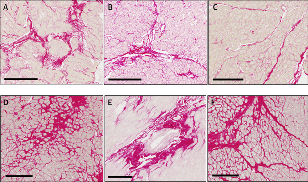
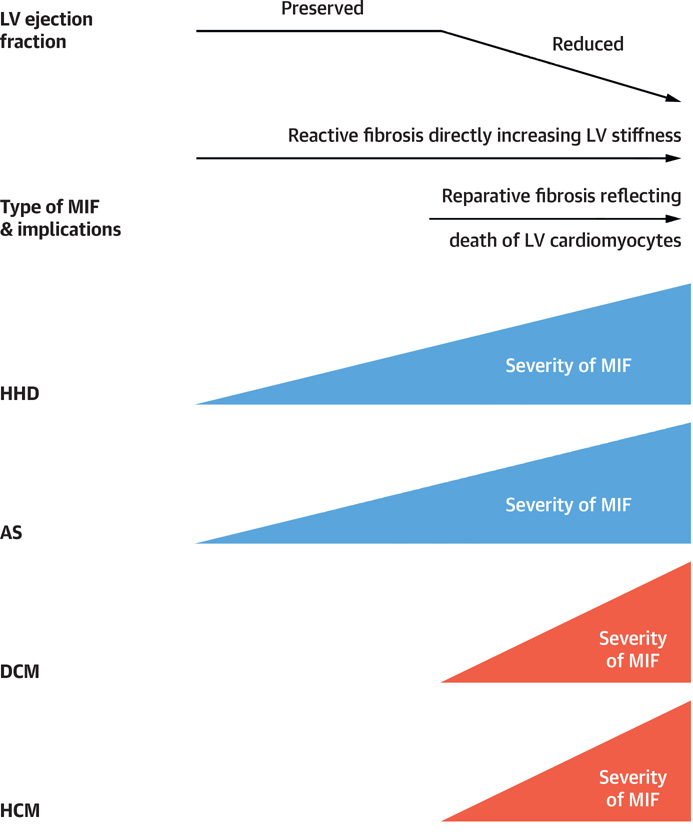
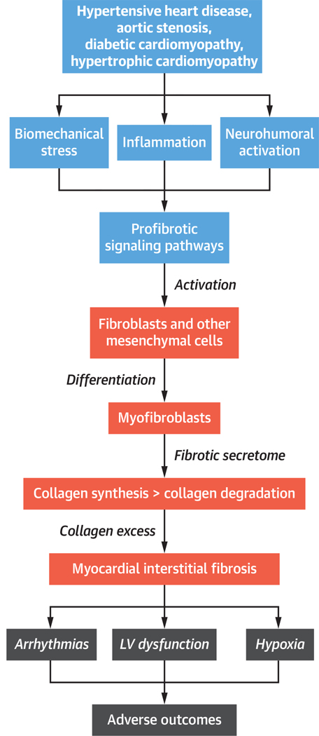
Myocardial interstitial fibrosis (MIF) is a histological hallmark of several cardiac diseases that alter myocardial architecture and function and are associated with progression to heart failure. MIF is a diffuse and patchy process, appearing as a combination of interstitial microscars, perivascular collagen fiber deposition, and increased thickness of mysial collagen strands. Although MIF arises mainly because of alterations in fibrillar collagen turnover leading to collagen fiber accumulation, there are also alterations in other nonfibrillar extracellular matrix components, such as fibronectin and matricellular proteins. Furthermore, in addition to an excess of collagen, qualitative changes in collagen fibers also contribute to the detrimental impact of MIF. In this part 3 of a 4-part JACC Focus Seminar, we review the evidence on the complex mechanisms leading to MIF, as well as its contribution to systolic and diastolic cardiac dysfunction and impaired clinical outcomes in patients with nonischemic heart disease.

Keywords: biomarker; collagen; fibroblast; heart failure; myocardial interstitial fibrosis.


Figures

FIGURE 1
FIGURE 1. Types of Fibrous Deposits in Myocardial Interstitial Fibrosis
Endomyocardial biopsy of (A-C) a patient with hypertensive heart disease and heart failure and (D-F) a patient with aortic valve stenosis showing myocardial interstitial fibrosis. Sections were stained with picrosirius red, and collagen deposits were identified in red as (A,D) microscars, (B,E) perivascular thick sheaths, and (C,F) thick bands deposited in the perimysium and the endomysium. Scale bars correspond to 200 μm.
FIGURE 2
FIGURE 2. Time Course of Changes in Severity of Myocardial Interstitial Fibrosis
Functional and clinical impact of myocardial interstitial fibrosis according to its histological type and clinical scenario. AS = aortic stenosis; DCM = diabetic cardiomyopathy; MIF = myocardial interstitial fibrosis; HCM = hypertrophic cardiomyopathy; HHD = hypertensive heart disease; LV = left ventricular.
FIGURE 3
FIGURE 3. Effect of Spironolactone on Patients With Heart Failure With Preserved Ejection Fraction
Peak early transmitral ventricular filling velocity to early diastolic tissue Doppler velocity ratio (E:e′) according to study treatment in tertiles of patients with low, intermediate, and high serum carboxy-terminal telopeptide of collagen type I to matrix metalloproteinase-1 ratio (CITP:MMP-1), which are equivalent to high, moderate, and low myocardial collagen cross-linking, respectively. Data are expressed as mean values and 95% confidence intervals at baseline and at 12 months in patients with heart failure with preserved ejection fraction treated with placebo or spironolactone. *p < 0.05 versus baseline. **p < 0.01 versus baseline. Reprinted with permission from Ravassa et al. (112).
CENTRAL ILLUSTRATION
CENTRAL ILLUSTRATION. The Process of Myocardial Interstitial Fibrosis in Nonischemic Heart Disease
Steps in the process of myocardial interstitial fibrosis, including associated clinical conditions, major mechanisms, and consequences. LV = left ventricular.

References

    1. Bowers SL, Banerjee I, Baudino TA. The extracellular matrix: at the center of it all. J Mol Cell Cardiol 2010;48:474–82. - PMC - PubMed
    1. Rienks M, Papageorgiou AP, Frangogiannis NG, Heymans S. Myocardial extracellular matrix: an ever-changing and diverse entity. Circ Res 2014;114:872–88. - PubMed
    1. Frangogiannis NG. The extracellular matrix in myocardial injury, repair, and remodeling. J Clin Invest 2017;127:1600–1612. - PMC - PubMed
    1. Brower GL, Gardner JD, Forman MF et al. The relationship between myocardial extracellular matrix remodeling and ventricular function. Eur J Cardiothorac Surg 2006;30:604–10. - PubMed
    1. Eckhouse SR, Spinale FG. Changes in the myocardial interstitium and contribution to the progression of heart failure. Heart Fail Clin 2012;8:7–20. - PMC - PubMed

Publication types