

Diagnostic Algorithm in Hirschsprung's Disease: Focus on Immunohistochemistry Markers
- PMID: 32354930
- PMCID: PMC7279842
- DOI: 10.21873/invivo.11913
Diagnostic Algorithm in Hirschsprung's Disease: Focus on Immunohistochemistry Markers
Abstract
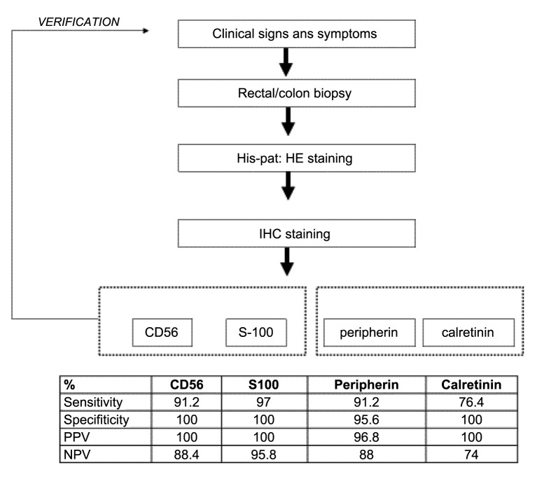
Background/aim: Hirschsprung disease (HD) is caused by the congenital absence of ganglion cells in the distal bowel (aganglionosis). Rectal biopsy is considered important for its diagnosis. The aim of this study was to apply immunohistochemical staining using a minimal set of antibodies and develop an algorithm that will assist in the diagnosis of HD.
Patients and methods: Rectal or colonic biopsies were performed in patients with HD (n=26) or patients treated for other bowel diseases (n=34). Immunohistochemical staining was performed for MAP1b, peripherin, S-100, calretinin, NSE, bcl-2 and CD56 proteins.
Results: Staining for CD56, S-100, peripherin and calretinin facilitated the identification of ganglion cells. The use of CD56 and S-100 antibodies together resulted in the highest rate of ganglion cell staining intensity (94%).
Conclusion: We propose a practical diagnostic algorithm with the application of CD56 and S-100 antibodies that can be used in clinical practice in children suspected of Hirschsprung's disease.
Keywords: CD56; Hirschsprung's disease; Immunohistochemistry; S-100; calretinin; ganglion cells; peripherin.
Copyright© 2020, International Institute of Anticancer Research (Dr. George J. Delinasios), All rights reserved.
Conflict of interest statement
The Authors have no conflicts of interest to disclose in regard to this study.
Figures

References
-
- Volpe A, Alaggio R, Midrio P, Iaria L, Gamba P. Calretinin, beta-tubulin immunohistochemistry, and submucosal nerve trunks morphology in Hirschsprung disease: Possible applications in clinical practice. J Pediatr Gastroenterol Nutr. 2013;57(6):780–787. doi: 10.1097/MPG.0b013e3182a934c7. - DOI - PubMed
MeSH terms
Substances
LinkOut - more resources
Full Text Sources
Research Materials