. 2020 Apr 30;10(1):7391.
doi: 10.1038/s41598-020-64464-9.

Potential toxicity of polystyrene microplastic particles

Affiliations

Potential toxicity of polystyrene microplastic particles

Jangsun Hwang et al. Sci Rep. .

Abstract

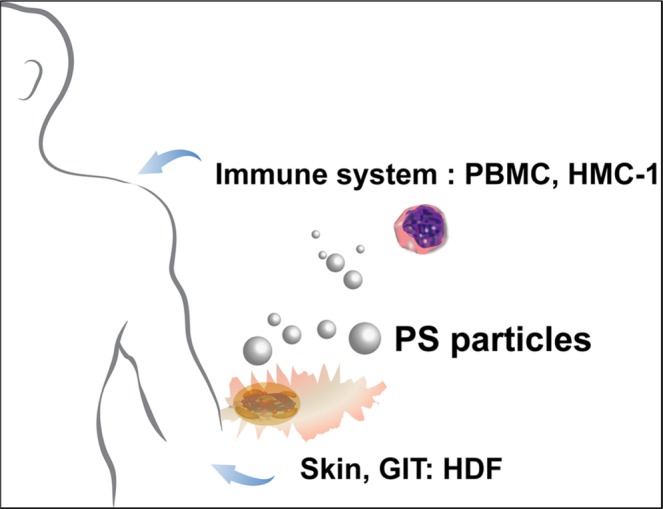
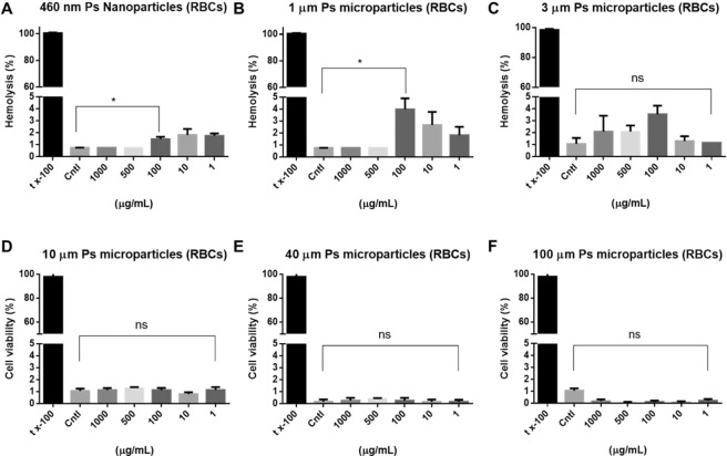
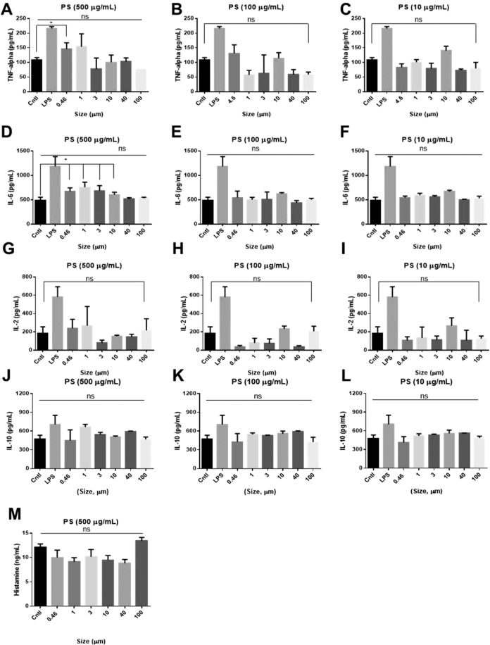
Environmental pollution arising from plastic waste is a major global concern. Plastic macroparticles, microparticles, and nanoparticles have the potential to affect marine ecosystems and human health. It is generally accepted that microplastic particles are not harmful or at best minimal to human health. However direct contact with microplastic particles may have possible adverse effect in cellular level. Primary polystyrene (PS) particles were the focus of this study, and we investigated the potential impacts of these microplastics on human health at the cellular level. We determined that PS particles were potential immune stimulants that induced cytokine and chemokine production in a size-dependent and concentration-dependent manner.


Conflict of interest statement

The authors declare no competing interests.

Figures

Figure 1
Figure 1
Illustration of the PS particle intake pathways of three cell lines. Human intake of PS particles from personal care products can occur via absorption through theskin. Intake can also occur through the ingestion of PS particles infood, food containers, drinking water, or biomedical products. We evaluated the potential of primary PS microparticles and nanoparticles tocause toxicity in humans based on the size and concentration of the particles in human cells.
Figure 2
Figure 2
SEM images and zeta potentials of PS particles. (A) 460 nm PS nanoparticles. (B) 1 µm PS particles. (C) 3 µm PS particles. (D) 10 µm PS particles. (E) 40 µm PS particles. (F) 100 µm PS particles (scale bar = 200 nm, 1 µm, 2 µm, 10 µm, and 20 µm). (G) Zeta potentials of the PS particles.
Figure 3
Figure 3
Cytotoxicity of PS particles. (A) 460 nm PS nanoparticles on HDFs. (B) 1 µm PS particles on HDFs. (C) 3 µm PS particles on HDFs. (D) 10 µm PS particles on HDFs. (E) 40 µm PS particles on HDFs. (F) 100 µm PS particles on HDFs. (G) 460 nm PS nanoparticles on PBMCs. (H) 3 µm PS particles on PBMCs. (I) 10 µm PS particles on PBMCs.
Figure 4
Figure 4
Confocal images of PS particles in cells. (A) Fluorescent images of 460 nm PS-FITC nanoparticles in PBMCs after DAPI staining (scale bar = 10 µm). Right: Z-section images. (B) Fluorescent images of 460 nm PS-FITC nanoparticles taken up by HDFs collected after DAPI staining (scale bar = 50 µm). Right: Z-section images.
Figure 5
Figure 5
Hemolysis of RBCs after contact with PS particles. (A) 460 nm PS nanoparticles. (B) 1 µm PS particles. (C) 3 µm PS particles. (D) 10 µm PS particles. (E) 40 µm PS particles. (F) 100 µm PS particles. 5% tx-100 served as the positive control. Cntl indicates no treatment. Absorbance was measured at 540 nm.
Figure 6
Figure 6
Cytokine profiles of TNF alpha, IL-2, IL-6, IL-10, and histamine. TNF-α secretion induced by PS particles of various sizes at concentrations of (A) 500 µg/mL, (B) 100 µg/mL, and (C) 10 µg/mL. IL-2 secretion induced by PS particles of various sizes at concentrations of (D) 500 µg/mL, (E) 100 µg/mL, and (F) 10 µg/mL. IL-6 secretion induced by PS particles of various sizes at concentrations of (G) 500 µg/mL, (H) 100 µg/mL, and (I) 10 µg/mL. IL-10 secretion induced by PS particles of various sizes at concentrations of (J) 500 µg/mL, (K) 100 µg/mL, and (L) 10 µg/mL. (M) Histamine profiles after treatment with 500 µg/mL PS particles of different sizes. Cntl: no treatment. LPS: 2.5 µg/mL.

References

    1. Law KL, Thompson RC. Microplastics in the seas. Science. 2014;345:144–145. doi: 10.1126/science.1254065. - DOI - PubMed
    1. Dauvergne P. The power of environmental norms: marine plastic pollution and the politics of microbeads. Environmental Politics. 2018;27:579–597. doi: 10.1080/09644016.2018.1449090. - DOI
    1. Fendall LS, Sewell MA. Contributing to marine pollution by washing your face: microplastics in facial cleansers. Marine pollution bulletin. 2009;58:1225–1228. doi: 10.1016/j.marpolbul.2009.04.025. - DOI - PubMed
    1. Napper IE, Bakir A, Rowland SJ, Thompson RC. Characterisation, quantity and sorptive properties of microplastics extracted from cosmetics. Marine Pollution Bulletin. 2015;99:178–185. doi: 10.1016/j.marpolbul.2015.07.029. - DOI - PubMed
    1. Gregory MR. Plastic ‘scrubbers’ in hand cleansers: a further (and minor) source for marine pollution identified. Marine pollution bulletin. 1996;32:867–871. doi: 10.1016/S0025-326X(96)00047-1. - DOI

Publication types