. 2020 Mar;8(6):350.
doi: 10.21037/atm.2020.02.103.

Instability and excessive mechanical loading mediate subchondral bone changes to induce osteoarthritis

Affiliations

Instability and excessive mechanical loading mediate subchondral bone changes to induce osteoarthritis

Jianxi Zhu et al. Ann Transl Med. 2020 Mar.

Abstract

Background: To assess the diversified effects of mechanical instability, excessive mechanical loading on subchondral bone remodeling. And to investigate the underlying cartilage degeneration and osteoarthritis (OA) progression in ipsilateral and contralateral knees, given that OA progression always affects joints bilaterally.

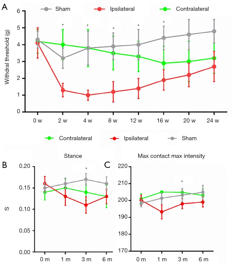
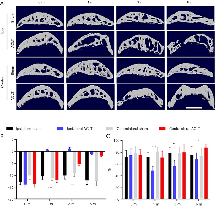
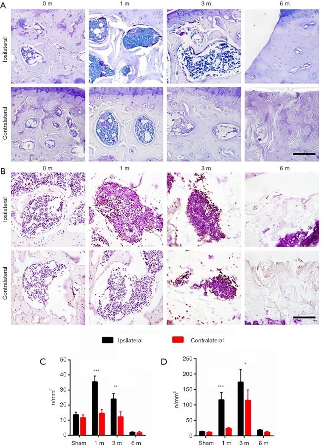
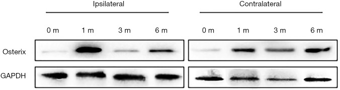
Methods: Anterior cruciate ligament transection (ACLT) of the left knee was used to induce OA in C57/B6 mice for 1, 3 and 6 months. Both left (ipsilateral) and right (contralateral) knees underwent micro-computerized tomography (micro-CT) scan and morphological analysis. The subchondral bone metabolism analysis by immunostaining of tartrate-resistant acid phosphatase (TRAP) and Osterix. Behavioral analyses including von Frey test and CatWalk gait analysis were also performed. Western blot analysis was performed to assess the signaling pathways involved in OA progression.

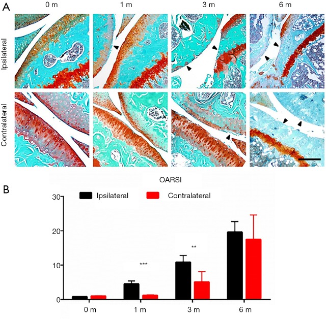
Results: Analyses showed that various changes in ipsilateral and contralateral knees lead to OA progression. Articular cartilage was rapidly destroyed on the ipsilateral side but was only gradually destroyed on the contralateral side. Micro-CT data showed a rapid decrease with a subsequent partial recovery of bone volume in the late stage on the ipsilateral side, while a gradual condensation of bone density was seen on the contralateral side. Immunostaining showed increased osteoclastic and osteoblastic activity in the early stage on the ipsilateral side, but only slight osteoblastic changes on the contralateral side. Behavioral analyses including von Frey and gait analysis showed that contralateral knees compensate ipsilateral mechanical loading, but also that this mechanism failed to work in the late stage.

Conclusions: Diversified mechanical loading properties lead to OA progression through different mechanisms of subchondral bone remodeling. Acute ACLT led to OA through bone density reduction, while the contralateral side developed OA gradually due to subchondral bone sclerosis.

Keywords: Osteoarthritis (OA); contralateral side; mechanical loading; subchondral bone.


Conflict of interest statement

Conflicts of Interest: YL serves as an unpaid section editor of Annals of Translational Medicine from Oct 2019 to Sep 2020. The other authors have no conflicts of interest to declare.

Figures

Figure 1
Figure 1
Ipsilateral and contralateral knees developed OA in distinct patterns in murine ACLT model. (A) Representative photos of Safranin O and fast green staining of ipsilateral (upper) an contralateral side of knee joints 0, 1, 3 and 6 months after ACLT; (B) statistical analysis, n=8 per group, statistical significance was determined by unpaired Student’s t test and all data were shown as a bar with means ± standard deviations. Scale bars, 500 µm. **, P<0.01, ***, P<0.001 compared with the sham operated group at different time points. ACLT, anterior cruciate ligament transection; OA, osteoarthritis.
Figure 2
Figure 2
Behavioral analysis reveals mechanical loading changes in OA progression of both knees. (A) von Frey analysis showing hind paw withdrawal threshold of sham operated mice, ipsilateral and contralateral side of mice in different time points after ACLT; CatWalk gait analysis including stance (B) and max contact max intensity (C) of sham operated mice, ipsilateral and contralateral side of mice in different time points after ACLT. n=8 per group, statistical significance was determined by multifactorial ANOVA and all data were shown as plots with means ± standard deviations. *, P<0.05 compared with the sham operated group at different time points. ACLT, anterior cruciate ligament transection; OA, osteoarthritis.
Figure 3
Figure 3
Differential subchondral bone architecture in ipsilateral and contralateral knees after ACLT. (A) Representative photos of CT 3D reconstruction of ipsilateral and contralateral tibial subchondral bone in different time points after ACLT; max contact max intensity (C) Statistical analysis of Tb Pf. (B) and BV/TV (C), n=8 per group. Statistical significance was determined by unpaired Student’s t test and all data were shown as a bar with means ± standard deviations. Scale bar, 2 mm; *, P<0.05, **, P<0.01, ***, P<0.001 compared with the sham operated group at different time points. ACLT, anterior cruciate ligament transection.
Figure 4
Figure 4
Differential osteoblastic and osteoclastic activities in ipsilateral and contralateral knees after ACLT. Representative photos of TRAP staining (A) and Osterix immunostaining (B) in ipsilateral and contralateral tibial subchondral bone in different time points after ACLT. statistical analysis of TRAP staining (C) and Osterix immunostaining (D), n=8 per group, statistical significance was determined by unpaired Student’s t test and all data were shown as a bar with means ± standard deviations. Scale bar, 20 µm; *, P<0.05, **, P<0.01, ***, P<0.001 compared with the sham operated group at different time points. ACLT, anterior cruciate ligament transection.
Figure 5
Figure 5
Western blot analysis of Osterix signal in ipsilateral and contralateral tibial subchondral bone in different time points after ACLT. ACLT, anterior cruciate ligament transection.

References

    1. Hootman JM, Helmick CG. Projections of US prevalence of arthritis and associated activity limitations. Arthritis Rheum 2006;54:226-9. 10.1002/art.21562 - DOI - PubMed
    1. Zhu J, Lei P, Hu Y. Intraarticular hyaluronate injection for knee osteoarthritis—reconsider the rationale. Ann Transl Med 2015;3:214. - PMC - PubMed
    1. Hunter DJ, Schofield D, Callander E. The individual and socioeconomic impact of osteoarthritis. Nat Rev Rheumatol 2014;10:437. 10.1038/nrrheum.2014.44 - DOI - PubMed
    1. Griffin TM, Guilak F. The role of mechanical loading in the onset and progression of osteoarthritis. Exerc Sport Sci Rev 2005;33:195-200. 10.1097/00003677-200510000-00008 - DOI - PubMed
    1. Read SJ, Dray A. Osteoarthritic pain: a review of current, theoretical and emerging therapeutics. Expert Opin Investig Drugs 2008;17:619-40. 10.1517/13543784.17.5.619 - DOI - PubMed