Randomized Controlled Trial
. 2020 May 1;143(5):1368-1382.
doi: 10.1093/brain/awaa052.

Corticospinal-motor neuronal plasticity promotes exercise-mediated recovery in humans with spinal cord injury

Affiliations
Randomized Controlled Trial

Corticospinal-motor neuronal plasticity promotes exercise-mediated recovery in humans with spinal cord injury

Hang Jin Jo et al. Brain. .

Abstract

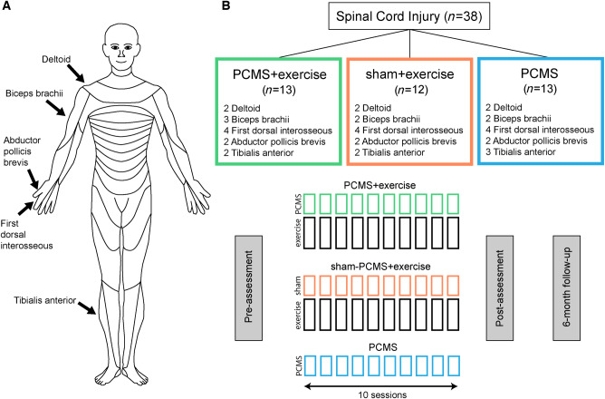
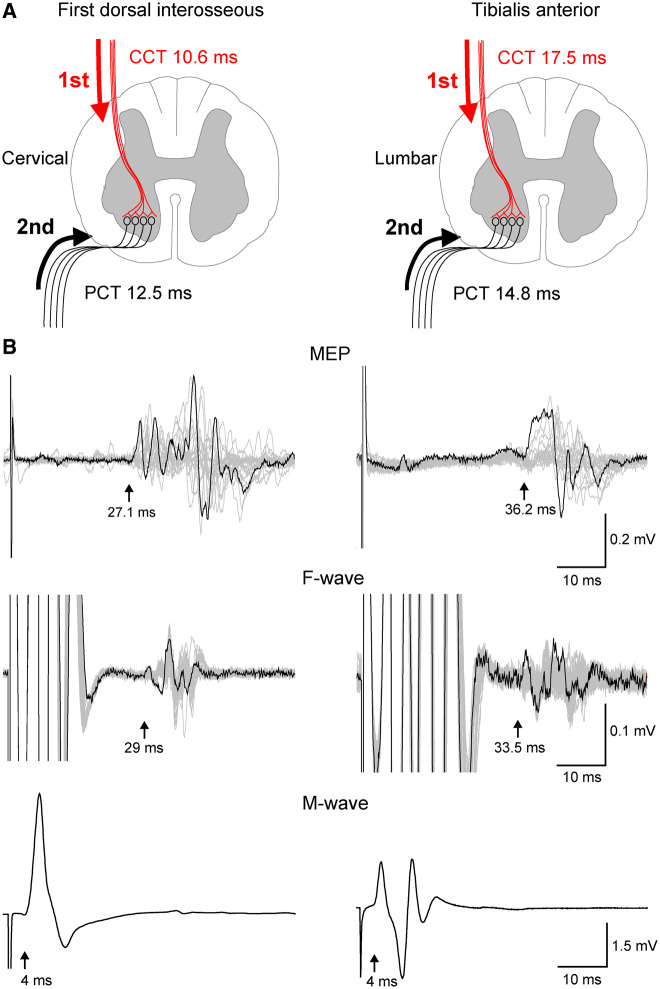
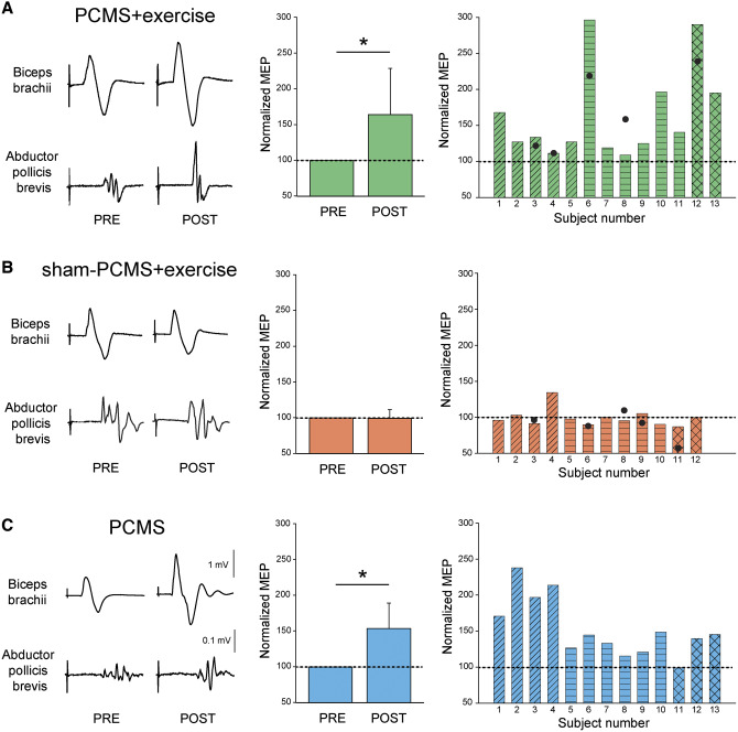
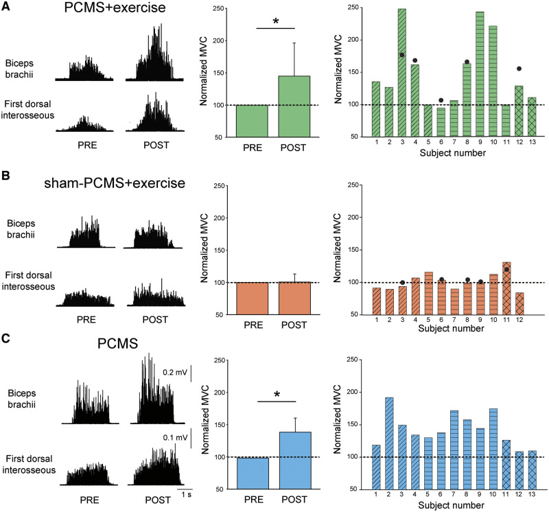
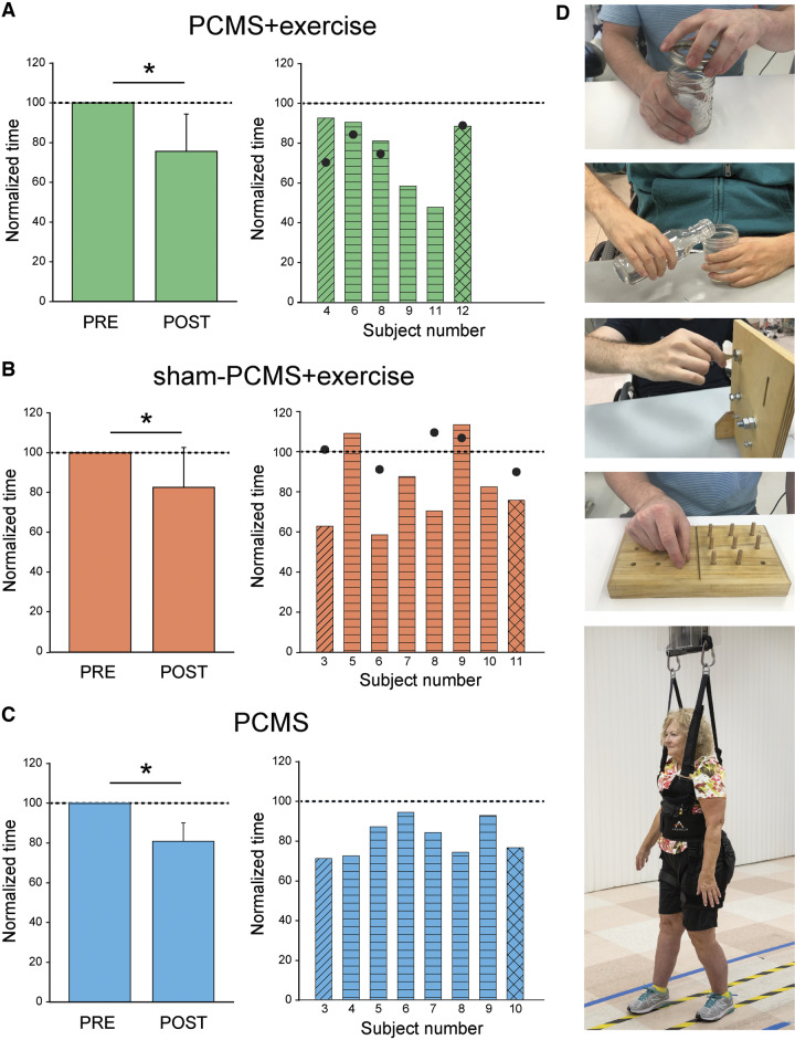
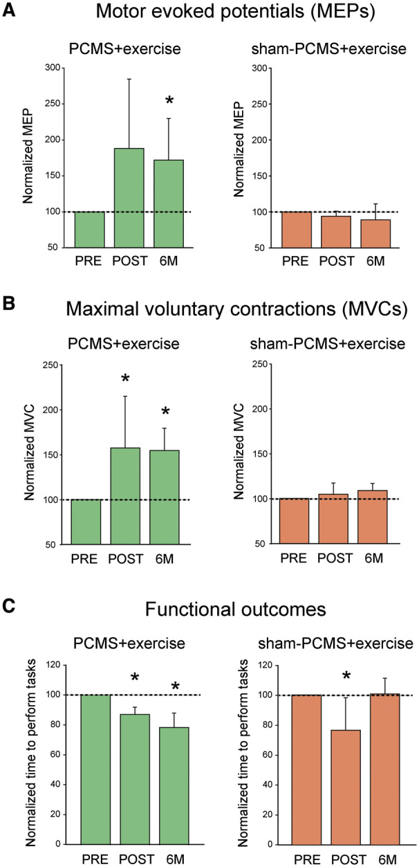
Rehabilitative exercise in humans with spinal cord injury aims to engage residual neural networks to improve functional recovery. We hypothesized that exercise combined with non-invasive stimulation targeting spinal synapses further promotes functional recovery. Twenty-five individuals with chronic incomplete cervical, thoracic, and lumbar spinal cord injury were randomly assigned to 10 sessions of exercise combined with paired corticospinal-motor neuronal stimulation (PCMS) or sham-PCMS. In an additional experiment, we tested the effect of PCMS without exercise in 13 individuals with spinal cord injury with similar characteristics. During PCMS, 180 pairs of stimuli were timed to have corticospinal volleys evoked by transcranial magnetic stimulation over the primary motor cortex arrive at corticospinal-motor neuronal synapses of upper- or lower-limb muscles (depending on the injury level), 1-2 ms before antidromic potentials were elicited in motor neurons by electrical stimulation of a peripheral nerve. Participants exercised for 45 min after all protocols. We found that the time to complete subcomponents of the Graded and Redefined Assessment of Strength, Sensibility and Prehension (GRASSP) and the 10-m walk test decreased on average by 20% after all protocols. However, the amplitude of corticospinal responses elicited by transcranial magnetic stimulation and the magnitude of maximal voluntary contractions in targeted muscles increased on overage by 40-50% after PCMS combined or not with exercise but not after sham-PCMS combined with exercise. Notably, behavioural and physiological effects were preserved 6 months after the intervention in the group receiving exercise with PCMS but not in the group receiving exercise combined with sham-PCMS, suggesting that the stimulation contributed to preserve exercise gains. Our findings indicate that targeted non-invasive stimulation of spinal synapses might represent an effective strategy to facilitate exercise-mediated recovery in humans with different degrees of paralysis and levels of spinal cord injury.

Keywords: exercise training; maximal voluntary contraction; motor evoked potentials; neuromodulation; spinal plasticity.


Figures

Figure 1
Figure 1
Experimental set-up. Diagrams showing muscles tested (A) and the study design (B). Thirty-eight individuals with chronic incomplete SCI were randomly assigned to 10 sessions of exercise combined with PCMS or sham-PCMS and PCMS.
Figure 2
Figure 2
Central (CCT) and peripheral (PCT) conduction time. Stimuli were timed to arrive at corticospinal-motor neuronal synapses by calculating CCT and PPT (A) using latencies from MEPs, F-waves, and M-waves (B). Traces shown from first dorsal interosseous and tibialis anterior muscles with latencies indicated by arrows.
Figure 3
Figure 3
MEPs. Raw MEP traces from six representative participants from biceps brachii and abductor pollicis brevis muscles before and after 10 sessions. Waveforms represent the average of 30 MEPs. Graphs show group (left) and individual (right) data for PCMS+exercise (A; n = 13) and sham-PCMS+exercise (B; n = 12), and PCMS (C; n = 13) groups. The x-axes of the left graphs show the time of measurements (PRE = pre-assessment; POST = post-assessment) and the y-axis shows the amplitude of MEPs as percentage of MEPs at pre-assessment. The x-axes of the right graphs show individual subjects and filled circles indicate the 6-month follow-up results. Data of participants included in the shoulder (transverse lines), hand (horizontal lines), and leg (crossed lines) blocks are shown for each intervention. Filled circles show individual MEP data in a subset of subjects at the 6-month follow-up after PCMS+exercise (n = 5) and sham-PCMS+exercise (n = 5). Scale bars shown for biceps brachii and first dorsal interosseous muscles are the same across participants. Error bars indicate SD, *P < 0.05.
Figure 4
Figure 4
MVCs. Rectified EMG traces during MVCs from six representative participants from biceps brachii and first dorsal interosseous muscles before and after 10 sessions. Graphs show group (left) and individual (right) data for PCMS+exercise (A; n = 13), sham-PCMS+exercise (B; n = 12), and PCMS (C; n = 13) groups. The x-axes of the left graphs show the time of measurements (PRE = pre-assessment; POST = post-assessment) and the y-axis shows the size of MVCs as percentage of MVCs at pre-assessment. The x-axes of the right graphs show individual subjects and filled circles indicate the 6-month follow-up results. Data of participants included in shoulder (transverse lines), hand (horizontal lines), and leg (crossed lines) block are shown for each intervention. Scale bars shown for biceps brachii and first dorsal interosseous muscles are the same across participants. Error bars indicate SDs, *P < 0.05.
Figure 5
Figure 5
Functional outcomes. Graphs show group (left) and individual (right) data for PCMS+exercise (A; n = 6), and sham-PCMS+exercise (B; n = 8), and PCMS (C; n = 8) groups. The x-axes show the time of measurements (PRE = pre-assessment; POST = post-assessment) and the y-axes show the time to perform tasks as percentage of the time at pre-assessment. The x-axes of the right graphs show individual subjects and filled circles indicate the 6-month follow-up results. (D) Tests involved subcomponents of the GRASSP and the 10-m walk tests. Data of participants included in the shoulder (transverse lines), hand (horizontal lines), and leg (crossed lines) block are shown for each intervention. Filled circles show individual functional outcomes in a subset of subjects at the 6-month follow-up after PCMS+exercise and sham-PCMS+exercise. Error bars indicate SDs, *P < 0.05.
Figure 6
Figure 6
Six-month follow-up results. Graphs show results after 10 sessions of PCMS+exercise (green bars) and sham-PCMS+exercise (orange bars) at the 6-month follow-up assessment. (A) MEPs (n = 5 for PCMS+exercise and 5 for sham-PCMS+exercise). The x-axis shows the time of assessments (PRE = pre-assessment; POST = post-assessment; 6M = 6-month follow-up assessment) and the y-axis shows the amplitude of MEPs as percentage of MEPs at pre-assessment (A), the MVCs as percentage of MVCs at pre-assessment (B), and the time to perform tasks as percentage of time at pre-assessment (C). Note that MEPs and MVCs increased after 10 sessions in the PCMS+exercise group and remained increased for 6 months compared with baseline but not in the sham-PCMS+exercise group. However, functional outcomes improved after 10 sessions of PCMS+exercise but did not persist 6 months later. Error bars indicate SDs, *P < 0.05.

References

    1. Angeli CA, Edgerton VR, Gerasimenko YP, Harkema SJ.. Altering spinal cord excitability enables voluntary movements after chronic complete paralysis in humans. Brain 2014; 137: 1394–409. - PMC - PubMed
    1. Ardestani MM, Henderson CE, Salehi SH, Mahtani GB, Schmit BD, Hornby TG.. Kinematic and neuromuscular adaptations in incomplete spinal cord injury after high- versus low-intensity locomotor training. J Neurotrauma 2019; 36: 2036–44. - PMC - PubMed
    1. Beekhuizen KS, Field-Fote EC.. Sensory stimulation augments the effects of massed practice training in persons with tetraplegia. Arch Phys Med Rehabil 2008; 89: 602–8. - PubMed
    1. Behrman AL, Ardolino EM, Harkema SJ.. Activity-based therapy: from basic science to clinical application for recovery after spinal cord injury. J Neurol Phys Ther 2017; 41 (Suppl 3): S39–S45. - PMC - PubMed
    1. Belci M, Catley M, Husain M, Frankel HL, Davey NJ.. Magnetic brain stimulation can improve clinical outcome in incomplete spinal cord injured patients. Spinal Cord 2004; 42: 417–9. - PubMed

Publication types