Review
. 2020 Dec;45(12):4012-4022.
doi: 10.1007/s00261-020-02547-x.

Delivering Clinical impacts of the MRI diagnostic pathway in prostate cancer diagnosis

Affiliations
Review

Delivering Clinical impacts of the MRI diagnostic pathway in prostate cancer diagnosis

Ivo G Schoots et al. Abdom Radiol (NY). 2020 Dec.

Abstract

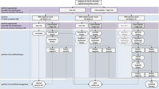
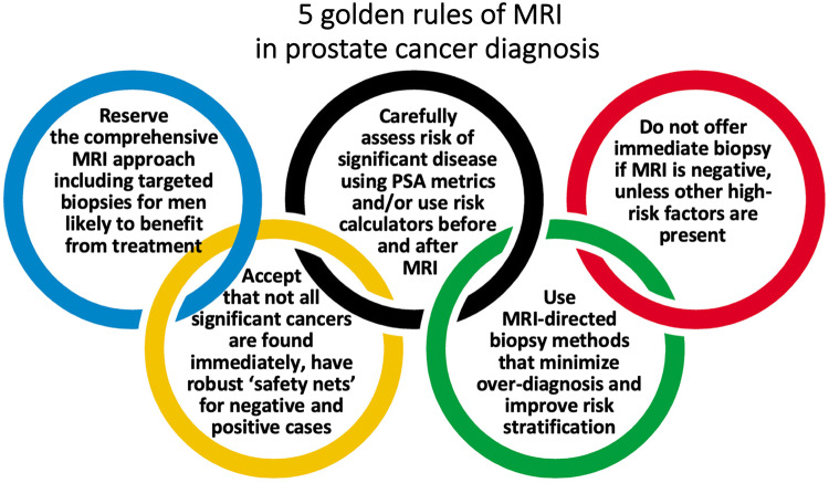
Pre-biopsy multiparametric MRI is now recommended by multiple guidelines, not only for men with persistent suspicion of prostate cancer after prior negative systematic biopsy, but also at initial screening before the first biopsy. The major benefit of pre-biopsy MRI in the diagnostic work-up is to promote individualized risk-adapted approaches for biopsy-decision management. Multiple MRI-directed diagnostic pathways can be conceived, with each approach having net-benefit trade-offs between benefits and harms, based on improved diagnostic yields of significant cancers and reduced biopsy testing and reduced detection of indolent prostate cancer. In this paper, we illustrate how clinical benefits can be maximized in men with MRI-negative and MRI-positive results, using the PI-RADS Multiparametric MRI and MRI-directed biopsy pathway. From a practice perspective, we emphasize five golden rules: (1) that multiparametric MRI approach including targeted biopsies be reserved for men likely to benefit from early detection and treatment of prostate cancer; (2) that there is a need to carefully assess risk of significant disease using PSA and clinical parameters before and after MRI; (3) do not offer immediate biopsy if the MRI is negative, unless other high-risk factors are present; (4) accept that not all significant cancers are found immediately and have robust 'safety nets' for men with negative MRI scans who avoid immediate biopsy and for positive MRI patients with negative or non-explanatory histology; and (5) use MRI-directed biopsy methods that minimize overdiagnosis and improve risk stratification.

Keywords: Biopsy; Magnetic resonance imaging (MRI); Multivariate risk prediction; Nomogram; Prostate cancer; Risk calculator; Risk stratification.


Conflict of interest statement

The authors have no potential conflict of interest to declare.

Figures

Fig. 1
Fig. 1
Flowchart of MRI pathway including patient risk stratification, when to perform biopsy. On the left suggested pre- and post-MRI risk stratification (purple) and related actions (blue), on the right the flowchart with actions (ovals) and results (rectangles). Identification of low-risk (light gray boxes) and intermediate-/high-risk men (darker gray boxes). Pathway preference (continuous lines) and alternative routes (dashed lines) are combined. *This route is more theoretical: a man with a PI-RADS 4 or 5 score at first diagnosis most likely will not be categorized in low risk, only after negative biopsies despite consistent PI-RADS 4 or 5 scores, as a results of MRI false positives. **This route with targeted and systematic biopsies can further be adapted to specific clinical scenarios. A man with a large PI-RADS 5 focus may already benefit from only MRI-directed targeted biopsy. A man with a small PI-RADS 4 focus may benefit from only MRI-directed focal saturation biopsy, sampling the suspected region and its penumbra. PSA prostate-specific antigen, MRI magnetic resonance imaging, GP general practitioner, MDT multidisciplinary team
Fig. 2
Fig. 2
A 69-year-old man with a PSA of 42 ng/ml. Digital rectal examination (DRE) was normal, and transrectal ultrasound (TRUS) showed a prostate volume of 231 ml, without any suspected hypo-echoic lesions found. The PSA density was 0.18 ng/ml2. The need to perform MRI and biopsy: This man’s risk cannot be estimated by using current risk calculators, due to prostate volume limitations (max. prostate volume of 110 cc). While PSA was very high, the PSA density was in the range of intermediate risk. A low pre-test probability would be expected. The MRI was negative (PI-RADS v2 score 2). Histopathology biopsy findings and treatment management: Systematic biopsies revealed no prostate cancer. This man is in the institutional follow-up protocol. Considerations: Despite the suggested high risk due to high PSA levels, men with large prostate volumes as a result of BPH are less likely to harbor significant cancers. For these men, systematic biopsies should be avoided to minimize overdiagnoses after a negative MRI
Fig. 3
Fig. 3
A 69-year-old man with a PSA of 6.9 ng/ml. Digital rectal examination (DRE) was normal, and transrectal ultrasound (TRUS) showed a prostate volume of 41 ml, without any hypo-echoic lesions found. The PSA density was 0.17 ng/ml2. The need to perform MRI and biopsy: Based on PSA and PSA density, a biopsy and a pre-biopsy MRI are both indicated. The MRI was negative (PI-RADS v2 score 2). Histopathology biopsy findings and treatment management: Systematic biopsies revealed no prostate cancer, only granulomatous inflammation on both sides. This man is in the institutional follow-up protocol. Considerations: In a man with a negative MRI with intermediate risk on clinical and PSA-density (≤ 0.15–0.20 ng/ml2) findings, deferral of immediate systematic biopsies can be considered with appropriate safety net protocol. Inflammation is a common cause of elevated serum PSA levels
Fig. 4
Fig. 4
a and b A 68-year-old man with a PSA of 17.2 ng/ml. Digital rectal examination (DRE) was normal, and transrectal ultrasound (TRUS) showed a prostate volume of 40 ml, without any hypo-echoic lesions found. The PSA density was 0.43 ng/ml2. The need to perform MRI and biopsy: Based on the PSA and PSA density, a biopsy and a pre-biopsy MRI are both indicated. However, based on the negative MRI findings a systematic biopsy should be discussed given the very elevated PSA density (≥ 0.20 ng/ml2). Histopathology biopsy findings and treatment: This man underwent systematic biopsies without cancer detection, and then urological follow-up. Follow-up management: At 4 years of follow-up, the PSA increased to 28.1 ng/ml, without DRE suspicion or volume change. Based on PSA and PSA density, a biopsy and a pre-biopsy MRI were again indicated, despite previous negative biopsies. Again, the MRI findings were negative, and a systematic biopsy again discussed, due to the elevated PSA density. Histopathology biopsy findings and treatment management: Systematic biopsies revealed one positive core out of seven left-sided cores, with a Gleason score of 3 + 3. Right-sided cores were negative for cancer. Patient preferred active surveillance outside protocol (high risk owing to single risk factor—PSA levels), instead of active treatment. Considerations: No significant cancer has been detected, despite very high levels of PSA. When the PSA level is high, the avoidance of systematic biopsies in MRI-negative men is considered unsafe as some significant cancers (mostly Gleason 3 + 4) can be present. A urological safety net should be in place for men avoiding immediate systematic biopsy after a negative MRI scan who remain at high suspicion
Fig. 5
Fig. 5
A 65-year-old man with a PSA of 10.6 ng/ml. Digital rectal examination (DRE) was normal, and transrectal ultrasound (TRUS) showed a prostate volume of 49 ml, without any hypo-echoic lesions found. The PSA density was 0.22 ng/ml2. The need to perform MRI and biopsy: Based on PSA, PSA density, a biopsy and a pre-biopsy MRI are both indicated. MRI identified a suspicious lesion of approximately 15 mm in the right transition zone, with substantial low ADC values and focal enhancement, without extraprostatic extension but with close proximity to the bladder neck. The total PI-RADS assessment category score was 5 (5/5/ +). Histopathology biopsy findings and treatment management: Biopsies revealed two positive cores out of three right-sided targeted cores with GS 3 + 3, without any cancer detection in five left-sided and five right-sided systematic biopsy cores. Patient preferred active surveillance outside clinical protocol (intermediate risk due to elevated PSA level > 10 ng/ml with visible tumor), instead of active treatment. Considerations: Use MRI findings as a roadmap to direct targeted biopsies. Large transition zone tumors may sometimes look very suspicious on imaging (PI-RADS 5) that on histologic evaluations have a GS 3 + 3 or GS 3 + 4. The clinical significance of these lesions is highly dependent on urologic preferences which should be understood by radiologists working in multidisciplinary teams
Fig. 6
Fig. 6
A 70-year-old man with a PSA of 11.1 ng/ml. Digital rectal examination (DRE) was normal, and transrectal ultrasound (TRUS) showed a prostate volume of 59 ml, without any hypo-echoic lesions found. The PSA density was 0.19 ng/ml2. The need to perform MRI and biopsy: Based on PSA and PSA density, a biopsy and a pre-biopsy MRI are both indicated. MRI identified a suspicious lesion of approximately 10 mm in the left peripheral zone, wedge-shaped with substantial low ADC values, with a total PI-RADS assessment category score 4 (4/4/ +). Histopathology biopsy findings and treatment management: Biopsies revealed two positive cores out of two left-sided targeted cores and two positive cores out of five right-sided systematic cores, all with GS 3 + 4 with cribriform growth present. Right-sided systematic biopsies also detected GS 3 + 3 in two out-of-five cores. Patient underwent robot-assisted radical prostatectomy with pT2a, GS3 + 4 with cribriform pathology with clear resection margins (R0). Considerations: When clinical and MRI findings are concordant, the likelihood of clinically significant cancers is increased. Targeted and systematic biopsies are required. Use MRI as a roadmap to direct biopsy cores. Note that cribriform pattern is underestimated by MRI. Note also that systematic cores are often more likely to be positive for cancer in sextants adjacent to MRI target lesions, hence the recommendation for focal saturation biopsies for small suspicious lesions
Fig. 7
Fig. 7
Five golden rules of MRI in prostate cancer diagnosis

References

    1. Padhani AR, Barentsz J, Villeirs G, Rosenkrantz AB, Margolis DJ, Turkbey B, Thoeny HC, Macura KJ, Tempany CM, Verma S, Weinreb JC. PI-RADS Steering Committee: The PI-RADS Multiparametric MRI and MRI-directed Biopsy Pathway. Radiology. 2019 doi: 10.1148/radiol.2019182946. - DOI - PMC - PubMed
    1. Mohler JL, Antonarakis ES, Armstrong AJ, D'Amico AV, Davis BJ, Dorff T, Eastham JA, Enke CA, Farrington TA, Higano CS, Horwitz EM, Hurwitz M, Ippolito JE, Kane CJ, Kuettel MR, Lang JM, McKenney J, Netto G, Penson DF, Plimack ER, Pow-Sang JM, Pugh TJ, Richey S, Roach M, Rosenfeld S, Schaeffer E, Shabsigh A, Small EJ, Spratt DE, Srinivas S, Tward J, Shead DA, Freedman-Cass DA. Prostate Cancer, Version 2.2019, NCCN Clinical Practice Guidelines in Oncology. J Natl Compr Canc Netw. 2019;17(5):479–505. doi: 10.6004/jnccn.2019.0023. - DOI - PubMed
    1. Bjurlin MA, Carroll PR, Eggener S, Fulgham PF, Margolis DJ, Pinto PA, Rosenkrantz AB, Rubenstein JN, Rukstalis DB, Taneja SS, Turkbey B. Update of the AUA Policy Statement on the Use of Multiparametric Magnetic Resonance Imaging in the Diagnosis, Staging and Management of Prostate Cancer. The Journal of urology. 2019;23(10):0000000000000617. - PMC - PubMed
    1. Mottet N, Bellmunt J, Bolla M, Briers E, Cumberbatch MG, De Santis M, Fossati N, Gross T, Henry AM, Joniau S, Lam TB, Mason MD, Matveev VB, Moldovan PC, van den Bergh RCN, Van den Broeck T, van der Poel HG, van der Kwast TH, Rouviere O, Schoots IG, Wiegel T, Cornford P (2020) EAU-ESTRO-ESUR-SIOG Guidelines on Prostate Cancer. https://uroweb.org/guideline/prostate-cancer/. - PubMed
    1. Schoots IG, Padhani AR, Rouviere O, Barentsz JO, Richenberg J. Analysis of Magnetic Resonance Imaging-directed Biopsy Strategies for Changing the Paradigm of Prostate Cancer Diagnosis. Eur Urol Oncol. 2019 doi: 10.1016/j.euo.2019.10.001. - DOI - PubMed