. 2020 Jun 1;96(6):fiaa079.
doi: 10.1093/femsec/fiaa079.

Effect of stevia on the gut microbiota and glucose tolerance in a murine model of diet-induced obesity

Affiliations

Effect of stevia on the gut microbiota and glucose tolerance in a murine model of diet-induced obesity

Sarah L Becker et al. FEMS Microbiol Ecol. .

Abstract

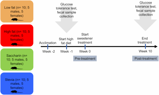
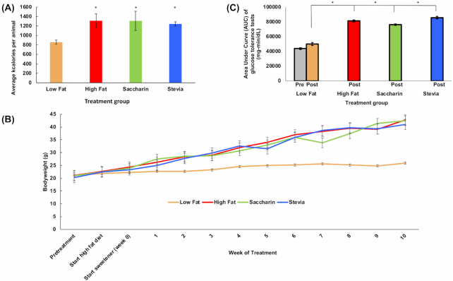
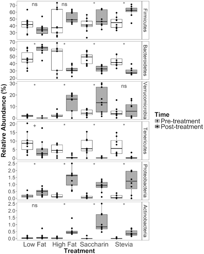
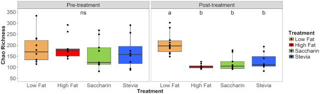
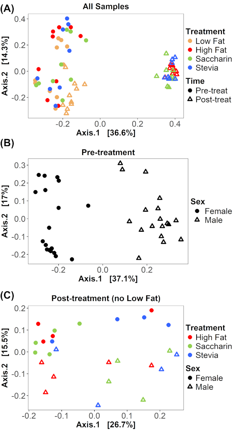
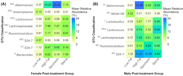
Artificial sweeteners have been shown to induce glucose intolerance by altering the gut microbiota; however, little is known about the effect of stevia. Here, we investigate whether stevia supplementation induces glucose intolerance by altering the gut microbiota in mice, hypothesizing that stevia would correct high fat diet-induced glucose intolerance and alter the gut microbiota. Mice were split into four treatment groups: low fat, high fat, high fat + saccharin and high fat + stevia. After 10 weeks of treatment, mice consuming a high fat diet (60% kcal from fat) developed glucose intolerance and gained more weight than mice consuming a low fat diet. Stevia supplementation did not impact body weight or glucose intolerance. Differences in species richness and relative abundances of several phyla were observed in low fat groups compared to high fat, stevia and saccharin. We identified two operational taxonomic groups that contributed to differences in beta-diversity between the stevia and saccharin groups: Lactococcus and Akkermansia in females and Lactococcus in males. Our results demonstrate that stevia does not rescue high fat diet-induced changes in glucose tolerance or the microbiota, and that stevia results in similar alterations to the gut microbiota as saccharin when administered in concordance with a high fat diet.

Keywords: Lactococcus; 16S rRNA; PERMANOVA; SIMPER; glucose tolerance; gut microbiota; saccharin.


Figures

Figure 1.
Figure 1.
Experimental design.
Figure 2.
Figure 2.
Physiological effects of stevia treatment. (A) Caloric intake over 10 week treatment indicates that animals on a high fat diet ate significantly more calories than those on a low-fat diet, and that addition of stevia or saccharin to the drinking water of mice on a high fat diet did not influence caloric intake. Graph displays aggregate data of both males and females. * P < 0.05 vs low fat. (B) Mice in the low fat group have significantly lower body weight than any of the mice in the experimental treatments (P < 0.0001). No significant difference was observed between groups on a high fat diet (high fat, saccharin, stevia). (C) Area under the curve (AUC) analysis of glucose tolerance tests in aggregate. All animals on a high fat diet showed elevated AUC when administered a glucose tolerance test (P < 0.0001 males, 0.003 females), and addition of NAS to the drinking water did not significantly affect the AUC vs high fat diet alone. Low fat was significantly lower than all other groups.
Figure 3.
Figure 3.
Mean relative abundance of phyla in samples categorized by treatment and time. Illustrated phyla have an average relative abundance >0.1% across all samples. Samples are categorized by treatment (horizontal) and colored by time, with pre-treatment samples as white and post-treatment samples as gray. Phyla are listed in order of highest to lowest average relative abundance and error bars represent the standard error. An increase in F/B ratio can be seen in all groups on a high fat diet in the post-treatment measurement. Annotations above the boxplots represent comparisons between pre- and post-treatment samples within each of the four treatment groups: an asterisk represents adjusted P < 0.05, a plus sign represents adjusted P = 0.05, and ‘ns’ represents adjusted P > 0.05 as compared by paired Wilcoxon ranked sums tests.
Figure 4.
Figure 4.
Chao richness is different in post-treatment samples. Samples from individual mice at pre- and post-treatment timepoints are separated by time (horizontal) and colored by treatment. Chao richness estimates the number of all OTUs in a sample. Pre-treatment samples were not significantly different (‘ns’; Kruskal–Wallis, P = 0.473). In post-treatment samples, samples labeled ‘a’ are significantly different from those labeled ‘b’. Post-treatment high fat, saccharin and stevia samples had significantly lower richness than that of low fat (Wilcoxon, adjusted P < 0.01). When comparing pre- and post-treatment samples within each treatment, richness decreased for high fat samples (Paired Wilcoxon, adjusted P = 0.016) but not for low fat (adjusted P = 0.432) or NAS treatments (saccharin adjusted P = 0.074, stevia adjusted P = 0.313).
Figure 5.
Figure 5.
Between-sample diversity is different between time, sex and treatment. Bray–Curtis dissimilarity is visualized on principal coordinate analysis (PCoA) ordinations. (A) All 72 samples from 36 individuals are displayed, with treatment represented by color and time (pre-treatment or post-treatment) represented by shape. With each treatment, all pre-treatment samples are significantly different from post-treatment samples (PERMANOVA, all adjusted P = 0.002). Notably, low fat post-treatment samples cluster distinctly from all other post-treatment samples. (B) Only 36 pre-treatment samples are displayed, with sex distinguished by shape. Sex is significantly different in pre-treatment samples (adjusted P = 0.001). (C) Only post-treatment samples from high fat diets (high fat, saccharin and stevia) are displayed (n = 26), with treatment represented by color and sex represented by shape. There is no significant difference between all high fat and all saccharin samples (adjusted P = 0.188) nor between all high fat and all stevia samples (adjusted P = 0.351). However, all saccharin and all stevia samples are significantly different (adjusted P = 0.039). When considering sex-driven differences, saccharin and stevia were significantly different in females (adjusted P = 0.048) but not males (adjusted P = 0.444).
Figure 6.
Figure 6.
Mean relative abundances of OTUs that significantly contribute to between-sample diversity. OTUs were identified using SIMPER and classification is shown for the most resolved taxonomic level. The heatmaps represent comparisons of treatment groups in post-treatment samples for females (A) and males (B). Annotations represent significant results for the following comparisons: A = low fat versus high fat, B = low fat versus saccharin, C = low fat versus stevia, D = saccharin versus stevia. Uppercase letters represent significance (adjusted P < 0.05) and lowercase letters represent a trend (0.10 > adjusted P > 0.05). Females had eight significant OTUs while males had seven trending OTUs. Almost all significant OTUs were identified in comparisons between low fat and high fat diet samples. Two OTUs contributed to differences between saccharin and stevia samples: one Lactococcus species and one Akkermansia species.

References

    1. Aghajanyan A, Movsisyan Z, Trchounian A. Antihyperglycemic and Antihyperlipidemic Activity of Hydroponic Stevia rebaudiana Aqueous Extract in Hyperglycemia Induced by Immobilization Stress in Rabbits. Biomed Res Int. 2017;2017:9251358. - PMC - PubMed
    1. Anton SD, Martin CK, Han Het al. .. Effects of stevia, aspartame, and sucrose on food intake, satiety, and postprandial glucose and insulin levels. Appetite. 2010;55:37–43. - PMC - PubMed
    1. Assaei R, Mokarram P, Dastghaib Set al. .. Hypoglycemic Effect of Aquatic Extract of Stevia in Pancreas of Diabetic Rats: PPARgamma-dependent Regulation or Antioxidant Potential. Avicenna journal of medical biotechnology. 2016;8:65–74. - PMC - PubMed
    1. Bailey CJ, Day C, Knapper JMet al. .. Antihyperglycaemic effect of saccharin in diabetic ob/ob mice. Br J Pharmacol. 1997;120:74–78. - PMC - PubMed
    1. Bian X, Tu P, Chi Let al. .. Saccharin induced liver inflammation in mice by altering the gut microbiota and its metabolic functions. Food Chem Toxicol. 2017;107:530–9. - PMC - PubMed

Publication types