Review
. 2020 Jun 1;219(6):e202002085.
doi: 10.1083/jcb.202002085.

Autophagosome biogenesis: From membrane growth to closure

Affiliations
Review

Autophagosome biogenesis: From membrane growth to closure

Thomas J Melia et al. J Cell Biol. .

Abstract

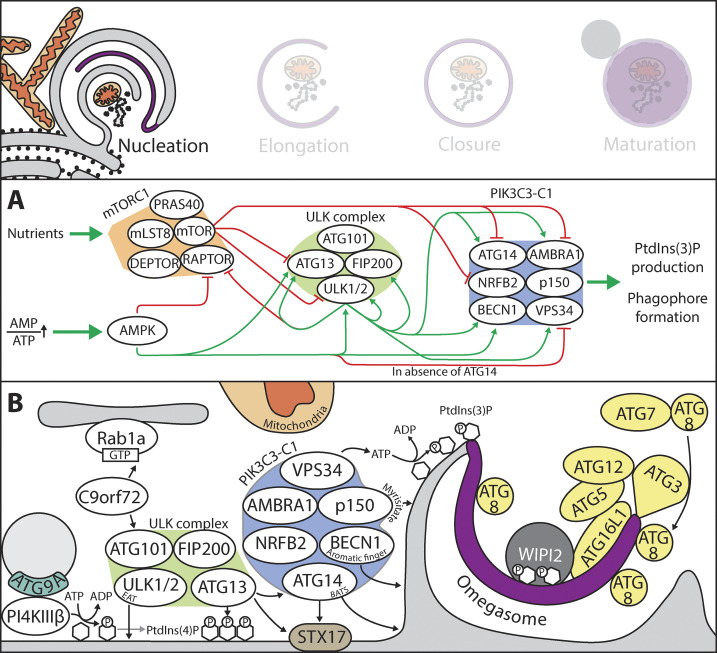
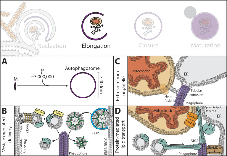
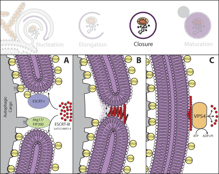
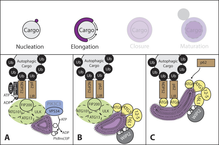
Autophagosome biogenesis involves de novo formation of a membrane that elongates to sequester cytoplasmic cargo and closes to form a double-membrane vesicle (an autophagosome). This process has remained enigmatic since its initial discovery >50 yr ago, but our understanding of the mechanisms involved in autophagosome biogenesis has increased substantially during the last 20 yr. Several key questions do remain open, however, including, What determines the site of autophagosome nucleation? What is the origin and lipid composition of the autophagosome membrane? How is cargo sequestration regulated under nonselective and selective types of autophagy? This review provides key insight into the core molecular mechanisms underlying autophagosome biogenesis, with a specific emphasis on membrane modeling events, and highlights recent conceptual advances in the field.


Figures

Figure 1.
Figure 1.
Overview of signaling events and protein–protein and protein–membrane interactions involved in phagophore nucleation. (A) Nutrient-rich conditions promote the activity of mTORC1, which inhibits autophagy by mTOR-mediated phosphorylations of the ULK complex (ULK1/2 and ATG13) and PIK3C3-CI (NRFB2, ATG14, and AMBRA1). In contrast, low energy status (high AMP-to-ATP ratio) causes activation of AMPK, which positively regulates autophagy by phosphorylation of the mTORC1 complex (Raptor), the ULK complex (ULK1 and ATG13), and PIK3C3-CI (BECN1 in the presence of ATG14 and VPS34 in the absence of ATG14). Activation of the ULK complex facilitates autophagy by autophosphorylation (ULK1, FIP200, and ATG13), inhibitory phosphorylation on mTORC1 (Raptor), and activating phosphorylations of the PIK3C3-CI (BECN1, VPS34, ATG14, and AMBRA1). (B) The autophagy-inducing signaling events described in A lead to membrane recruitment of the ULK complex. This is promoted by its interaction with C9orf72 (Rab1 effector) and dependent on the EAT domain of ULK1 and the N-terminus of ATG13. The latter interacts with acidic phospholipids, including PtdIns(4)P, generated by the PI4KIIIβ, which interacts with ATG9, a transmembrane protein important for phagophore elongation (see Fig. 2). The ULK complex stabilizes the PIK3C3-CI through direct interactions between ATG14 and ATG13. ATG14 also interacts with the ER-resident protein STX17 at ER–mitochondria contact sites. PIK3C3-CI membrane binding is further mediated by ATG14 (N-terminal cysteine-rich domain and a PtdIns(3)P binding BATS domain), BECN1 (aromatic finger), and p150 (N-terminal myristate). Generation of PtdIns(3)P by the PIK3C3-CI facilitates recruitment of the PtdIns(3)P effector protein WIPI2 that promotes ATG8 conjugation to PE through recruitment of the ATG12-ATG5-ATG16L1 complex, an E3 of the ATG8 conjugation machinery. Lipidated ATG8 proteins can function as a scaffold for core autophagy machinery components and as membrane attachment sites for autophagic cargo receptors (see Fig. 4). The phagophore membrane is indicated in purple.
Figure 2.
Figure 2.
Model of suggested mechanisms involved in phagophore elongation. (A) A rough estimate shows that ∼3 million lipids could be required to produce an autophagosome of 400 nm (see Box 2). Three distinct mechanisms for delivery of lipids for phagophore elongation have been proposed: vesicle-mediated delivery, membrane extrusion from pre-existing organelles, and protein-mediated lipid transport. (B) For vesicle-mediated delivery, ATG9- and ATG16L1-positive vesicles formed from recycling endosomes (dependent on SNX18, DNM2, and adaptor proteins) and COPII vesicles from ER exit sites (ERES) and ERGIC have been implicated in phagophore elongation. (C) For membrane extrusion from preexisting organelles, tubular extrusions from the ER and mitochondria have been proposed to form the expanding phagophore. (D) For protein-mediated lipid transport, illustrated for ATG2A and GRAMD1A, ATG2A acts as a lipid tunnel with little or no lipid specificity, while GRAMD1A functions as a cholesterol transfer protein.
Figure 3.
Figure 3.
Autophagosome closure is facilitated by the ESCRT machinery. (A) ESCRT-I components are recruited to the phagophore by an unknown mechanism, followed by recruitment of the filament-forming ESCRT-III components CHMP2A and CHMP4B. In yeast, Atg17 (FIP200) interacts with the ESCRT-III subunit Snf7 (CHMP4), indicating a role for the ULK complex in recruitment of ESCRT-III for phagophore closure. (B) ESCRT-III polymerization leads to filament formation, bringing the leading edge of the phagophore into close apposition to allow membrane fission. (C) Recruitment of the AAA-ATPase VPS4 resolves the fission process and facilitates depolymerization of the ESCRT-III filament structure. ATG8 proteins are also implicated in phagophore elongation and closure, but the mechanisms involved are not clear.
Figure 4.
Figure 4.
Hypothetical model for de novo autophagosome formation during selective autophagy. (A) Autophagy receptors (p62 and NDP52) bound to selective cargo interact directly with FIP200, leading to recruitment of the ULK complex as well as VPS34. TBK1-mediated phosphorylation of NDP52 stimulates the NDP52–FIP200 interaction. The p62–FIP200 interaction requires the LIR domain in p62. (B) PtdIns(3)P production by VPS34 causes recruitment of WIPI2 and the ATG8 conjugation machinery, leading to ATG8 lipidation. (C) The p62-FIP200 binding can be outcompeted by binding of p62 to ATG8, which facilitates further recruitment of autophagy receptors and expansion of the autophagic membrane tightly around the selective substrate.

References

    1. Abeliovich H., Dunn W.A. Jr., Kim J., and Klionsky D.J.. 2000. Dissection of autophagosome biogenesis into distinct nucleation and expansion steps. J. Cell Biol. 151:1025–1034. 10.1083/jcb.151.5.1025 - DOI - PMC - PubMed
    1. Agudo-Canalejo J., and Knorr R.L.. 2019. Formation of Autophagosomes Coincides with Relaxation of Membrane Curvature. Methods Mol. Biol. 1880:173–188. 10.1007/978-1-4939-8873-0_10 - DOI - PubMed
    1. Alemu E.A., Lamark T., Torgersen K.M., Birgisdottir A.B., Larsen K.B., Jain A., Olsvik H., Øvervatn A., Kirkin V., and Johansen T.. 2012. ATG8 family proteins act as scaffolds for assembly of the ULK complex: sequence requirements for LC3-interacting region (LIR) motifs. J. Biol. Chem. 287:39275–39290. 10.1074/jbc.M112.378109 - DOI - PMC - PubMed
    1. Andrejeva G., Gowan S., Lin G., Wong Te Fong A.L., Shamsaei E., Parkes H.G., Mui J., Raynaud F.I., Asad Y., Vizcay-Barrena G., et al. . 2019. De novo phosphatidylcholine synthesis is required for autophagosome membrane formation and maintenance during autophagy. Autophagy. 10.1080/15548627.2019.1659608 - DOI - PMC - PubMed
    1. Antonioli M., Albiero F., Nazio F., Vescovo T., Perdomo A.B., Corazzari M., Marsella C., Piselli P., Gretzmeier C., Dengjel J., et al. . 2014. AMBRA1 interplay with cullin E3 ubiquitin ligases regulates autophagy dynamics. Dev. Cell. 31:734–746. 10.1016/j.devcel.2014.11.013 - DOI - PubMed

Publication types

MeSH terms