. 2020 May 18;7(3):ENEURO.0047-20.2020.
doi: 10.1523/ENEURO.0047-20.2020. Print 2020 May/Jun.

Behavioral Characterization of dmrt3a Mutant Zebrafish Reveals Crucial Aspects of Vertebrate Locomotion through Phenotypes Related to Acceleration

Affiliations

Behavioral Characterization of dmrt3a Mutant Zebrafish Reveals Crucial Aspects of Vertebrate Locomotion through Phenotypes Related to Acceleration

Ana Del Pozo et al. eNeuro. .

Abstract

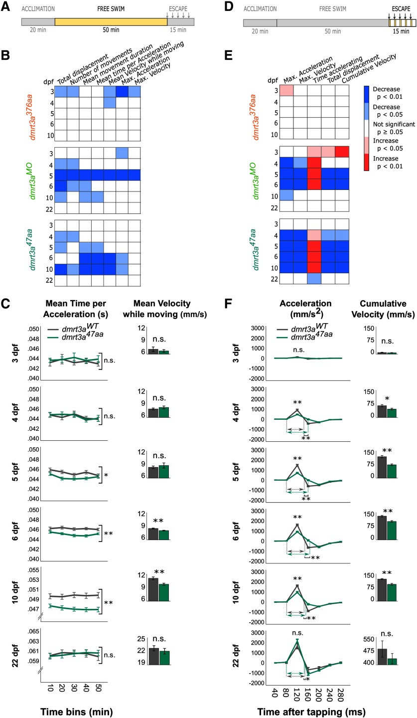
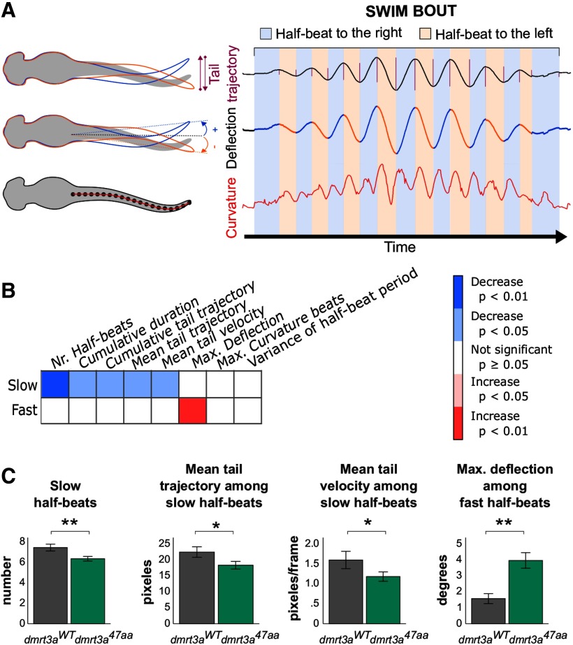
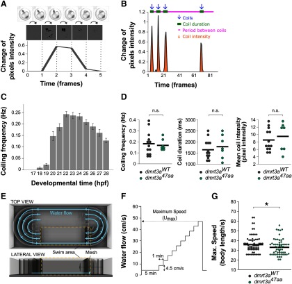
Vertebrate locomotion is orchestrated by spinal interneurons making up a central pattern generator. Proper coordination of activity, both within and between segments, is required to generate the desired locomotor output. This coordination is altered during acceleration to ensure the correct recruitment of muscles for the chosen speed. The transcription factor Dmrt3 has been proposed to shape the patterned output at different gaits in horses and mice. Here, we characterized dmrt3a mutant zebrafish, which showed a strong, transient, locomotor phenotype in developing larvae. During beat-and-glide swimming, mutant larvae showed fewer and shorter movements with decreased velocity and acceleration. Developmental compensation likely occurs as the analyzed behaviors did not differ from wild-type at older larval stages. However, analysis of maximum swim speed in juveniles suggests that some defects persist within the mature locomotor network of dmrt3a mutants. Our results reveal the pivotal role Dmrt3 neurons play in shaping the patterned output during acceleration in vertebrates.

Keywords: Danio rerio; central pattern generator; gait; locomotion; spinal cord; wt1.


Figures

Figure 1.
Figure 1.
Description of the zebrafish models used. A, Schematic structure of the predicted Dmrt3a protein, showing the DNA binding domain (DM) and the Dmrt3 family domain (DMA). Lighter shade represents missing amino acids compared with the wild-type form of Dmrt3a. B, Western blot for Dmrt3a (47 kDa) and β-actin (42 kDa) protein at 3 dpf in dmrt3aWT, dmrt3a47aa, and dmrt3a376aa as well as 2 and 3 dpf in dmrt3aMO. C, Alignment of dmrt3a cDNA partial sequences between dmrt3aWT (top) and the CRISPR/Cas9 generated mutant dmrt3a47aa(bottom). The fragment shows the proximity to the 5 bp deletion (—) where the asterisk (*) represents the premature stop codon generated and the underlined sequence indicates the sgRNA target. This figure is extended in Extended Data Figure 1-1.
Figure 2.
Figure 2.
Comparative locomotor analyses of the three zebrafish models (dmrt3a376aa, dmrt3aMO, and dmrt3a47aa) in relation to their dmrt3aWT. A, D, Schematic of experimental protocol indicates whether data below belong to the free swimming (A) or to the escape response (D); arrows designate timing of taps. B, E, Heat maps to visualize statistical differences among dmrt3a376aa, dmrt3aMO, and dmrt3a47aa in relation to their dmrt3aWT during 1–6 and 10 dpf over the 50 min of free swimming (B) and over the 280 ms of induced escape responses (E). Increase (red) and decrease (blue) of parameters in mutant larvae and morphants compared with dmrt3aWT. C, Mean time per acceleration of dmrt3a47aa and their dmrt3aWT are represented in 10 min bins during free swimming for a dynamic visualization over the trial. However, statistics are performed on the whole trial. Inset, Bar graphs show the mean velocity while moving over the trial. F, Acceleration dynamic of dmrt3a47aa and their dmrt3aWT during escape response in 40 ms bins. Inset, bar graphs show cumulative velocity over the escape. Dashed horizontal arrows indicate the duration of the acceleration phase (in ms) for each group when they are significantly different. All data are plotted as the mean ± SEM. *p < 0.05. **p < 0.01. n.s. - not statistically significant. Statistical data from this figure are shown in Extended Data Figure 2-1.
Figure 3.
Figure 3.
Tail kinematics in dmrt3a47aa animals. A, Representative trace of a larva, illustrating the definition of parameters analyzed, such as tail trajectory, deflection, and curvature. B, Heat map to visualize statistical differences between 6 dpf dmrt3a47aa and dmrt3aWT, during both slow and fast half-beats, separately. The increase (red) and decrease (blue) of parameters in mutant larvae are compared with dmrt3aWT. C, Most relevant parameters where dmrt3a47aa differed from dmrt3aWT (i.e., the number of slow half-beats, mean tail trajectory among the slow half-beats, mean tail trajectory, and mean tail velocity among the slow half-beats, and the maximum deflection among the fast half-beats). All data are plotted as the mean ± SEM. *p < 0.05. **p < 0.01. Statistical data from this figure are shown in Extended Data Figure 3-1.
Figure 4.
Figure 4.
Locomotor activity of dmrt3a47aa embryos and juveniles. A, Coil movement detection by quantifying pixel change from frame to frame. B, A representative analysis and extraction of parameters. Detected coils (blue arrows), coiling duration (green bars), intercoil duration (pink line), and coil intensity (orange area) were quantified for each individual. C, Coil frequency (Hz) in wild-type zebrafish from 17 to 28 hpf. D, Coiling frequency (Hz), duration (ms), and coil intensity (pixel intensity) of dmrt3a47aa and dmrt3aWT. n.s., p ≥ 0.05. E, Schematic of swim tunnel used for analysis in juveniles: top and lateral views. F, Experimental protocol applied to determine maximum speed (Umax). G, Maximum swim speed in body lengths per second reached by the dmrt3a47aa and dmrt3aWT. Individual data are represented by dots, and the mean is indicated by a horizontal line. *p < 0.05. n.s. - not statistically significant. Statistical data from this figure are shown in Extended Data Figure 3-1.
Figure 5.
Figure 5.
Expression of dmrt3a and related genes in dmrt3a47aa and dmrt3aWT. A, Relative expression of dmrt3a mRNA from 1 to 5 dpf. B, Schematic of a 3 dpf larva where relevant structures are labeled in purple [i.e., hindbrain (HB), spinal cord (SC), olfactory bulb (OB), and kidney (K)]. C–J, mRNA pattern expression at 3 dpf. Top and lateral views: images of dmrt3a47aa and dmrt3aWT represent the average expression of 5 larvae. Merged color images below show the expression from dmrt3aWT in green and from dmrt3a47aa in magenta. Top view: C, dmrt3a expression; D, dmrt1a expression; E, dmrt2a expression; F, wt1a expression. Lateral view: G, dmrt3a expression; H, dmrt1a expression; I, dmrt2a expression; J, wt1a expression. Abbreviations highlight relevant labeling. This figure is extended in Extended Data Figure 5-1.

References

    1. Allalou A, Wu Y, Ghannad-Rezaie M, Eimon PM, Yanik MF (2017) Automated deep-phenotyping of the vertebrate brain. Elife 6:e23379 10.7554/eLife.23379 - DOI - PMC - PubMed
    1. Ampatzis K, Song J, Ausborn J, El Manira A (2014) Separate microcircuit modules of distinct V2a interneurons and motoneurons control the speed of locomotion. Neuron 83:934–943. 10.1016/j.neuron.2014.07.018 - DOI - PubMed
    1. Andersson LS, Larhammar M, Memic F, Wootz H, Schwochow D, Rubin C-J, Patra K, Arnason T, Wellbring L, Hjälm G, Imsland F, Petersen JL, McCue ME, Mickelson JR, Cothran G, Ahituv N, Roepstorff L, Mikko S, Vallstedt A, Lindgren G, et al. (2012) Mutations in DMRT3 affect locomotion in horses and spinal circuit function in mice. Nature 488:642–646. 10.1038/nature11399 - DOI - PMC - PubMed
    1. Berg EM, Björnfors ER, Pallucchi I, Picton LD, El Manira A (2018) Principles governing locomotion in vertebrates: lessons from zebrafish. Front Neural Circuits 12:73. 10.3389/fncir.2018.00073 - DOI - PMC - PubMed
    1. Björnfors ER, El Manira A (2016) Functional diversity of excitatory commissural interneurons in adult zebrafish. Elife 5:e18579 10.7554/eLife.18579 - DOI - PMC - PubMed

Publication types

LinkOut - more resources