Review
. 2020 May 1;147(9):dev182279.
doi: 10.1242/dev.182279.

Neuronal and glial regulation of CNS angiogenesis and barriergenesis

Affiliations
Review

Neuronal and glial regulation of CNS angiogenesis and barriergenesis

Saptarshi Biswas et al. Development. .

Abstract

Neurovascular pathologies of the central nervous system (CNS), which are associated with barrier dysfunction, are leading causes of death and disability. The roles that neuronal and glial progenitors and mature cells play in CNS angiogenesis and neurovascular barrier maturation have been elucidated in recent years. Yet how neuronal activity influences these processes remains largely unexplored. Here, we discuss our current understanding of how neuronal and glial development affects CNS angiogenesis and barriergenesis, and outline future directions to elucidate how neuronal activity might influence these processes. An understanding of these mechanisms is crucial for developing new interventions to treat neurovascular pathologies.

Keywords: Angiogenesis; Astrocytes; Basement membrane; Blood-brain barrier; Blood-retina barrier; Light response; Müller glia; Neuroglial progenitors; Neuronal activity; Neurovascular unit; Radial glia; Retinal waves.


Conflict of interest statement

Competing interestsThe authors declare that they have no relevant affiliation or competing financial interests with any organization or entity having a financial interest or conflict with materials discussed in the manuscript.

Figures
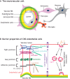
Fig. 1.
Fig. 1.
Interactions between various cell types in the neurovascular unit are crucial for the formation of neurovascular barriers. (A) In the CNS, vascular components, including endothelial cells (light gray), endothelial cell-derived basement membrane (endothelial BM; yellow) and mural cells [such as pericytes (pink)], together with astrocytes (purple), astrocyte-derived BM (astrocyte BM; green), neurons (gray) and microglia (orange) interact anatomically to form the neurovascular unit (NVU). The boxed area is presented in greater detail in B. (B) A schematic of the cell biological mechanisms that contribute to the neurovascular barrier properties of CNS endothelial cells. Tight junctions, formed by claudins (dark blue), occludin (maroon) and zonula occludens (light blue), limit the movement of small molecules between endothelial cells, thereby forming a paracellular barrier. Tight junction proteins interact with the cytoskeleton (F-actin, green) via zonula occludens proteins (light blue). Adherens junctions, which are formed by VE-cadherin (dark gray) as well as α-catenin (dark brown), β-catenin (medium brown) and γ-catenin (light brown), are crucial for cell-cell interactions and shear stress sensing. Endothelial cells communicate with each other via connexin-regulated gap junctions (dark green). Low rates of caveolin 1 (Cav1, pink)-dependent receptor (dashed black)-mediated transcytosis prevent trafficking of larger molecules and antibodies within CNS endothelial cells, establishing a transcellular barrier. Finally, CNS endothelial cells also contain specific active and passive transporters (purple) to facilitate the movement of nutrients between the blood and the brain/retina.
Fig. 2.
Fig. 2.
Developmental timelines for neurogenesis, gliogenesis, angiogenesis and barriergenesis in the brain and retina. (A,B) Developmental timelines (i) and schematic illustrations (ii) of the development of neurons, glial cells and blood vessels in the mouse cerebral cortex (A) and retina (B). (Ai) Neurogenesis in the cerebral cortex begins at E11, whereas gliogenesis starts at E13 (oligodendrocyte formation) and E18.5 (astrocyte formation). Neurogenesis is completed by P8-P10, whereas gliogenesis persists for prolonged periods in postnatal development corresponding to the expansion and maturation of astrocytes. Cortical angiogenesis also spans both the embryonic and postnatal stages of development, and is completed by P25. The initiation of BBB maturation [establishment of both paracellular (blue) and transcellular (pink) properties] starts soon after angiogenesis. However, by birth, both the paracellular and transcellular barrier properties of brain endothelial cells are mature. Dotted lines indicate the initiation and maturation of the endothelial barrier properties or the start of differentiation and appearance of mature astrocytes and oligodendrocytes. (Aii) Neurogenesis in the cortex occurs in an inside-out fashion (gray arrow), whereas angiogenesis occurs in an outside-in fashion (red arrow). Mature astrocytes ensheath blood vessels to ensure the maintenance of the BBB. (Bi) Amacrine cells, cones, ganglion cells and horizontal cells are born during the embryonic phase in the retina, whereas bipolar cells and Müller glia are born during the postnatal phase. Rod development occurs throughout both phases. Astrocytes migrate into the retina from E18.5 until P6. Growth of the superficial plexus (P1-P8) follows an astrocyte template, over the inner limiting membrane. Sprouts from the superficial vessels, guided by Müller glia, then form the deep (P8-P12) and intermediate (P12-P17) plexuses. The transcellular BRB matures by P10, whereas the paracellular barrier does not mature until P18. Upward arrows indicate the peak of development for each process. Dotted lines indicate the initiation and maturation of BRB properties in retinal endothelial cells. (Bii) Schematic diagram shows the relationship between distinct neuronal cell types and distinct vascular plexuses in the retina. Ganglion cell bodies reside in the ganglion cell layer, which is vascularized by the superficial plexus. Amacrine, bipolar, horizontal and Müller cell bodies reside in the inner nuclear layer, which is vascularized by the intermediate and deep vascular plexuses. Photoreceptors occupy the outer nuclear layer, which makes contacts with the deep vascular plexus.
Fig. 3.
Fig. 3.
Developmental timeline of neuronal activity and BBB/BRB maturation in the mouse CNS. Developmental timeline of synaptogenesis, neural activity and BBB/BRB maturation in the mouse cerebral cortex (A) and retina (B). (A) Synaptogenesis (light-gray bar) in the mouse cortex begins around E13.5 (E16.5 is when activity-dependent synaptic pruning begins), whereas activity-dependent synaptic pruning (dark-gray bar) begins at E16.5 and peaks at birth. Both processes continue through P21. The transcellular barrier properties of the BBB (blue curve) in the cortex do not completely mature until E16-E17, and the paracellular BBB properties (pink curve) do not mature until birth, when the transendothelial electrical resistance (TEER) is highly increased. (B) Between E16 and P0, retinal waves (gray bars) are propagated by gap junctions (stage I). From P0-P10, these waves are cholinergic in nature (stage II), whereas between P10-P14 they switch to being glutamatergic (stage III). Most neuronal activity (dark-gray bars) in the mouse retina after P14 is light mediated (i.e. photoreceptor dependent), although some reports suggest that photoreceptor activity occurs as early as P10. Moreover, the light response by intrinsically photosensitive melanopsin ganglion cells occurs throughout this period (E16-adult). By P10, the endothelial transcellular BRB (pink curve) is mature throughout all retinal vascular beds. In contrast, the paracellular BRB (blue curve) properties of the retinal vasculature gradually arise until P18.
Fig. 4.
Fig. 4.
The main neuronal and glial-derived signals that influence angiogenesis and barriergenesis. Schematic representation of the signaling interactions between endothelial cells and other cell types in the cerebral cortex (A) and the retina (B). (A) In the cerebral cortex, radial glia, neuroglial progenitors and neurons secrete VEGFA. Radial glia and neuroglial progenitors also secrete several Wnt ligands; radial glia also secrete TGFβ. Astrocytes produce Ang1 and sonic hedgehog (Shh), and these same cells in the adult cortex secrete VEGFA during injury. Finally, Cajal-Retzius cells express reelin. Pro-angiogenic processes are depicted in dark green, whereas anti-angiogenic processes are depicted in light green. (B) In the retina, neuronal cell types and Müller glia secrete VEGFA, which facilitates vascular growth in distinct layers. Ganglion cells, amacrine cells and bipolar cells secrete reelin, whereas amacrine cells and bipolar cells produce Slit2, both of which promote angiogenesis. Depending on tissue physiology (such as severe ischemia), ganglion cells also secrete semaphorin 3A and semaphorin 3E (Sema3A and Sema3E), which inhibit retinal vascular growth. Müller glia secrete norrin, which promotes retinal angiogenesis and BRB formation. In contrast, Müller glia-derived TGFβ inhibits endothelial cell proliferation and vascular branching. TGFβ1 derived from resting microglia inhibits endothelial cell proliferation, while Wnt5a and Wnt11 produced by these cells negatively regulate vascular branching in the deep plexus. In contrast, activated microglia induce endothelial cell proliferation, likely by secreting tumor necrosis factor α (TNFα). Pro-angiogenic factors are depicted in dark green, whereas anti-angiogenic factors are depicted in light green.

References

    1. Alvarez J. I., Dodelet-Devillers A., Kebir H., Ifergan I., Fabre P. J., Terouz S., Sabbagh M., Wosik K., Bourbonniere L., Bernard M. et al. (2011). The Hedgehog pathway promotes blood-brain barrier integrity and CNS immune quiescence. Science 334, 1727-1731. 10.1126/science.1206936 - DOI - PubMed
    1. Armulik A., Genové G., Mäe M., Nisancioglu M. H., Wallgard E., Niaudet C., He L., Norlin J., Lindblom P., Strittmatter K. et al. (2010). Pericytes regulate the blood-brain barrier. Nature 468, 557-561. 10.1038/nature09522 - DOI - PubMed
    1. Arnold T. D., Ferrero G. M., Qiu H., Phan I. T., Akhurst R. J., Huang E. J. and Reichardt L. F. (2012). Defective retinal vascular endothelial cell development as a consequence of impaired integrin alphaVbeta8-mediated activation of transforming growth factor-beta. J. Neurosci. 32, 1197-1206. 10.1523/JNEUROSCI.5648-11.2012 - DOI - PMC - PubMed
    1. Arnold T. D., Niaudet C., Pang M.-F., Siegenthaler J., Gaengel K., Jung B., Ferrero G. M., Mukouyama Y. S., Fuxe J., Akhurst R. et al. (2014). Excessive vascular sprouting underlies cerebral hemorrhage in mice lacking alphaVbeta8-TGFbeta signaling in the brain. Development 141, 4489-4499. 10.1242/dev.107193 - DOI - PMC - PubMed
    1. Bader B. L., Rayburn H., Crowley D. and Hynes R. O. (1998). Extensive vasculogenesis, angiogenesis, and organogenesis precede lethality in mice lacking all alpha v integrins. Cell 95, 507-519. 10.1016/S0092-8674(00)81618-9 - DOI - PubMed

Publication types