Review
. 2020 May 1;5(47):eaaz9631.
doi: 10.1126/sciimmunol.aaz9631.

Regulatory T cells in skin injury: At the crossroads of tolerance and tissue repair

Affiliations
Review

Regulatory T cells in skin injury: At the crossroads of tolerance and tissue repair

Ian C Boothby et al. Sci Immunol. .

Abstract

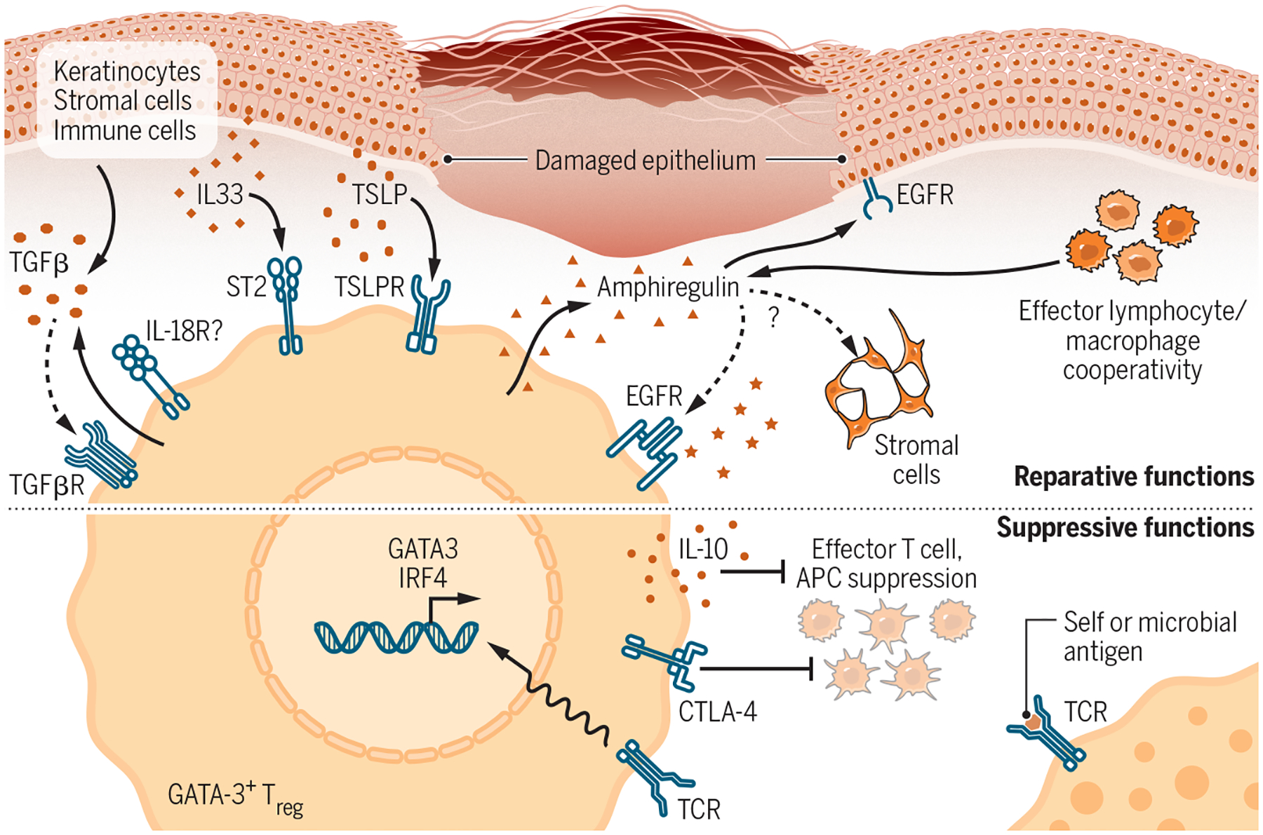
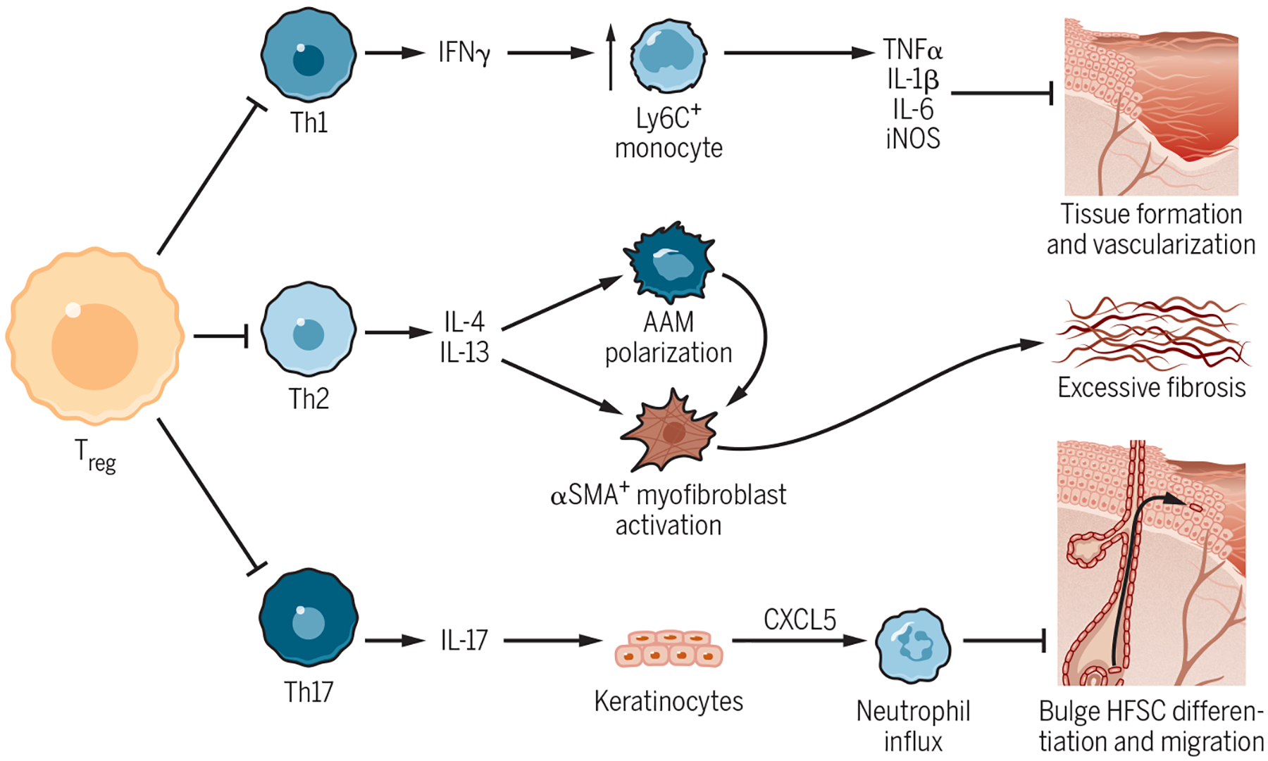
Skin injury is a highly inflammatory process that is carefully regulated to mitigate tissue damage and allow for proper barrier repair. Regulatory T cells (Tregs) are crucial coordinators of the immune response to injury in several organs. Here, we review the emerging role of Tregs in facilitating skin repair after injury. We focus on recently discovered interactions between lymphocytes and nonhematopoietic cells during wound healing and discuss how these interactions are regulated both by "classical" suppressive mechanisms of Tregs and by "nonclassical" reparative Treg functions.


Figures

Figure 1-
Figure 1-. Progression of Acute Wound Healing in Skin.
(A) Immediately after wounding, a fibrin clot forms to plug the damaged tissue. (B) For several days after injury, local release of tissue damage signals leads to the recruitment of neutrophils and monocytes/macrophages from circulation to prevent infection and phagocytose dead cells and debris. Recent evidence suggests that nearby tissue-resident lymphocytes can also mobilize to wound margins during this time period. (C) As inflammation resolves, keratinocytes at the wound margins proliferate, migrate, and differentiate to re-epithelialize the injury site and restore barrier integrity. Granulation tissue forms in the wound bed and is vascularized by angiogenesis of nearby blood vessels. Myofibroblasts accumulate in granulation tissue and contract to draw the wound closed. The influx of lymphocytes into the wound reaches its peak, and macrophage polarization shifts from pro-inflammatory to pro-reparative states. (D) After the wound closes, cellularity of the wound bed decreases and a scar forms through a combination of collagen deposition, cross-linking, and remodeling. This process strengthens the wound site at the expense of regenerating normal dermis.
Figure 2-
Figure 2-. Participation of Type 2 and Type 3 Lymphocytes in Cutaneous Tissue Repair.
Left: The type 2 immune response to skin injury is triggered by release of alarmins such as TSLP, IL-18, and IL-33 from damaged keratinocytes, endothelial cells, and stromal cells. Locally resident type 2 lymphocytes such as ILC2s sense these tissue damage signals and produce IL-4, IL-13, and amphiregulin in response. These cytokines promote wound closure by activating wound-contracting myofibroblasts, both directly by signaling to nearby fibroblasts and indirectly by inducing alternative activation of macrophages (AAM). Type 2 cytokines are also capable of driving keratinocyte proliferation and therefore may promote re-epithelialization. Right: Type 3 lymphocytes are abundant in skin and respond to both microbial ligands and tissue damage signals released by keratinocytes and myeloid cells, including IL-1β and IL-23. IL-17 and IL-22 produced by these cells act directly on keratinocytes, resulting in a two-pronged tissue protective response. First, antimicrobial immunity is bolstered by neutrophil recruitment and antimicrobial peptide production. Secondly, these cytokines reinforce the epidermal barrier by driving keratinocyte proliferation to cover injury sites at the expense of keratinocyte maturation. Solid arrows, known interactions; Dotted arrows, likely interactions based on data from other tissues and contexts.
Figure 3-
Figure 3-. Reparative and Suppressive Effector Mechanisms of Type 2-Polarized Tregs.
Skin bears a high proportion of type 2-polarized Tregs programmed by Th2-associated transcription factors such as GATA-3 and IRF4 that both confer tissue-reparative functions. Top: GATA-3+ Tregs in skin express receptors for alarmins such as TSLP, IL-33, and possibly IL-18 that are released upon tissue damage, enabling them to sense local injury. IL-18 and IL-33 can both stimulate Treg production of the reparative cytokine amphiregulin independent of TCR stimulation. Amphiregulin (AREG) drives keratinocyte proliferation and may promote regeneration of stromal populations in wounded skin. Skin Tregs additionally express both TGF-β and TGFβR, although the Treg-specific function of each is not entirely clear. Many of these reparative functions are shared by effector lymphocyte and macrophage populations. Bottom: In addition to conferring direct tissue-reparative function to Tregs, GATA-3 and IRF4 also bolster adaptive Treg functions in part by stabilizing FoxP3 expression. These “classical” Treg functions are linked to TCR stimulation and include production of the regulatory cytokine IL-10 and suppression of costimulation on antigen presenting cells by Treg-expressed CTLA-4. Solid arrows, known interactions; Dotted arrows, likely interactions based on data from other tissues and contexts.
Figure 4-
Figure 4-. Regulation of Wound Healing by Classical Treg Suppressive Functions.
(A) Early after injury, Tregs suppress excessive production of IFN-γ by Th1 cells and thus restrain the accumulation of pro-inflammatory Ly6C+ monocytes/macrophages. These cells impede the transition to the proliferative phase of healing by secreting highly inflammatory cytokines such as TNF-α, IL-1β, and IL-6. (B) Impaired Treg control of type 2 immunity leads to Th2 accumulation in wounded skin and excessive production of IL-4 and IL-13. These cytokines drive collagen deposition by activating pro-fibrotic α-SMA+ myofibroblasts and alternatively activated macrophages. Treg depletion consequently worsens cutaneous fibrosis. (C) After epidermal barrier damage, Tregs promote re-epithelialization by hair follicle stem cells (HFSCs) by suppressing excessive type 3 immunity. Loss of Tregs results in uncontrolled Th17 and neutrophil accumulation, which inhibit the differentiation and migration of HFSCs from the hair follicle bulge into the interfollicular epidermis.

References

    1. Eming SA, Hammerschmidt M, Krieg T & Roers A Interrelation of immunity and tissue repair or regeneration. Semin. Cell Dev. Biol 20, 517–527 (2009). - PubMed
    1. Ali N & Rosenblum MD Regulatory T cells in skin. Immunology (2017). doi:10.1111/imm.12791 - DOI - PMC - PubMed
    1. Blanpain C & Fuchs E Epidermal homeostasis: a balancing act of stem cells in the skin. Nat Rev Mol Cell Bio 10, nrm2636 (2009). - PMC - PubMed
    1. Cotsarelis G, Sun TT & Lavker RM Label-retaining cells reside in the bulge area of pilosebaceous unit: Implications for follicular stem cells, hair cycle, and skin carcinogenesis. Cell (1990). doi:10.1016/0092-8674(90)90696-C - DOI - PubMed
    1. Wang X et al. Principles and mechanisms of regeneration in the mouse model for wound-induced hair follicle neogenesis. Regeneration 2, 169–181 (2015). - PMC - PubMed

Publication types