. 2020 May 19;117(20):10976-10982.
doi: 10.1073/pnas.1917125117. Epub 2020 May 1.

Acoustofluidic sonoporation for gene delivery to human hematopoietic stem and progenitor cells

Affiliations

Acoustofluidic sonoporation for gene delivery to human hematopoietic stem and progenitor cells

Jason N Belling et al. Proc Natl Acad Sci U S A. .

Abstract

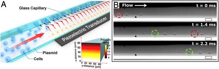
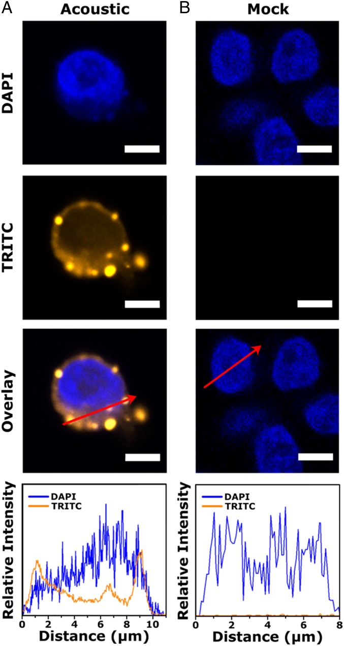
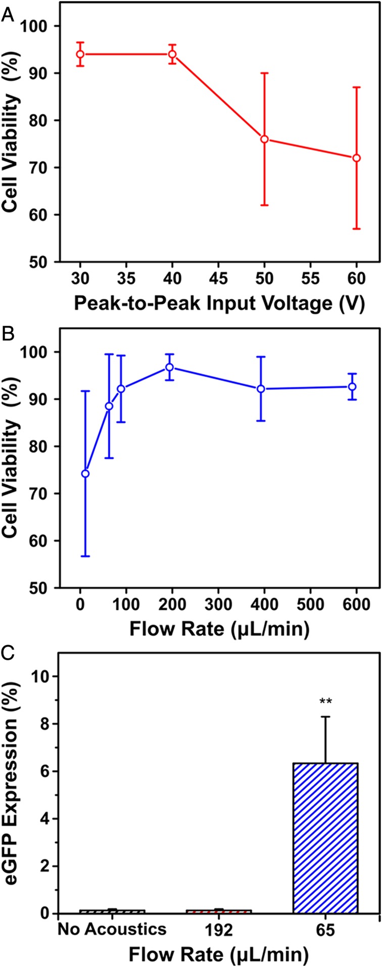
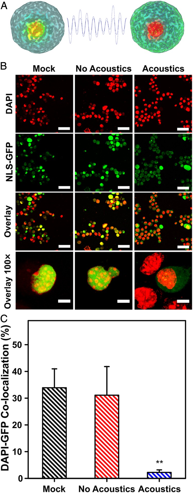
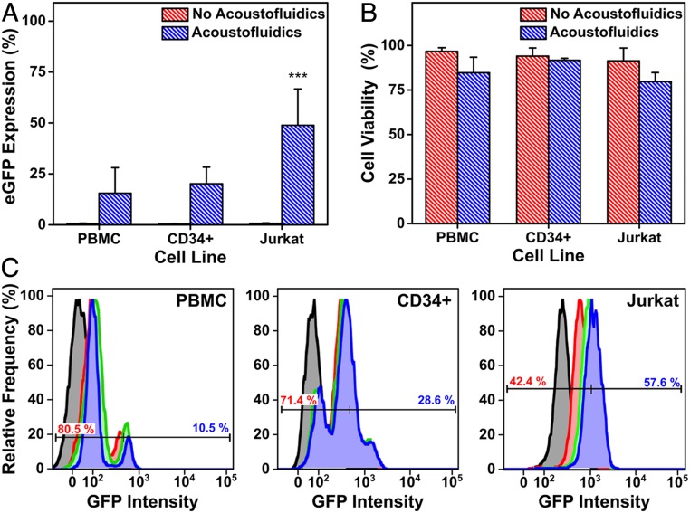
Advances in gene editing are leading to new medical interventions where patients' own cells are used for stem cell therapies and immunotherapies. One of the key limitations to translating these treatments to the clinic is the need for scalable technologies for engineering cells efficiently and safely. Toward this goal, microfluidic strategies to induce membrane pores and permeability have emerged as promising techniques to deliver biomolecular cargo into cells. As these technologies continue to mature, there is a need to achieve efficient, safe, nontoxic, fast, and economical processing of clinically relevant cell types. We demonstrate an acoustofluidic sonoporation method to deliver plasmids to immortalized and primary human cell types, based on pore formation and permeabilization of cell membranes with acoustic waves. This acoustofluidic-mediated approach achieves fast and efficient intracellular delivery of an enhanced green fluorescent protein-expressing plasmid to cells at a scalable throughput of 200,000 cells/min in a single channel. Analyses of intracellular delivery and nuclear membrane rupture revealed mechanisms underlying acoustofluidic delivery and successful gene expression. Our studies show that acoustofluidic technologies are promising platforms for gene delivery and a useful tool for investigating membrane repair.

Keywords: acoustofluidics; gene therapy; hematopoietic stem cells; intracellular delivery.


Conflict of interest statement

Competing interest statement: P.S.W., S.J.J., A.Z.S., and J.N.B. are inventors on US and international patent applications filed by the Regents of the University of California relating to the acoustofluidic platform.

Figures

Fig. 1.
Fig. 1.
(A) Schematic of the device components and application, where target cells undergo acoustofluidic treatment via flow through a glass capillary over a piezoelectric transducer and are collected at the outlet. (Inset) Simulated acoustic pressure amplitude of the aqueous medium in the glass capillary showing minimum pressure presents at the wall farthest from the piezoelectric transducer at an excitation frequency of 3.3 MHz. (B) Sequential images taken with a high-speed camera at 0, 1.4, and 2.2 ms. Jurkat cells are observed to localize against a capillary wall and are pushed forward by laminar flow. Colored circles are used to track cells moving through the capillary. (Scale bars, 50 μm.)
Fig. 2.
Fig. 2.
Confocal laser scanning micrographs for (A) acoustic-treated and (B) untreated Jurkat cells. Line profiles (red arrows) of 4′,6-diamidino-2-phenylindole (DAPI) and TRITC channels show fluorescence signal of Cy3-labeled DNA at the cell membrane, cytosol, and nucleus for acoustic-treated cells. The overlays show the two images above, combined with ImageJ software. Scale bars are 10 μm.
Fig. 3.
Fig. 3.
(A) Cell viability as a function of the applied peak-to-peak input voltage to the piezoelectric transducer with a constant flow rate of 192 μL/min. (B) Cell viability as a function of flow rate through the glass capillary with a constant input voltage of 40 V peak-to-peak. (C) eGFP in Jurkat cells 24 h postacoustofluidic delivery of an eGFP-expression plasmid. Protein expression is plotted as a function of flow rate and compared to a no acoustics control flowed at 65 μL/min. All cell viability measurements were assessed through trypan blue staining. Data are expressed as mean and SD for n = 3. Significance is determined using a one-way ANOVA and a Tukey means comparison test (**P < 0.01).
Fig. 4.
Fig. 4.
(A) A schematic of MEFs green fluorescent protein fused to a nuclear localization signal (NLS-GFP) and acoustofluidic treatment inducing nuclear membrane rupture, resulting in dispersed NLS-GFP throughout the cell cytosol. (B) Confocal laser scanning micrographs of MEFs showing colocalization events of NLS-GFP and DAPI signals in the untreated (mock), no acoustics, and acoustics-treated samples. The MEFs are virally transduced to express GFP at their nuclei (green) and are stained postacoustofluidic treatment with DAPI to label the cell nuclei (red). Colocalization of GFP and DAPI signals (yellow) are shown in the overlay, and lack thereof is evidence of nuclear membrane rupture. (Scale bars, upper three rows, 50 μm; bottom row, 10 μm.) (C) Quantification of colocalized DAPI and GFP signals for mock, no acoustic, and acoustic-treated cells. Data are expressed as mean and SD for n = 3 and significance is determined using a one-way ANOVA and Tukey’s mean comparison test (**P < 0.01). Colocalization % of DAPI and GFP signals are normalized to 60 cells for each condition.
Fig. 5.
Fig. 5.
eGFP expression (A) and cell viability (B) 72 h postacoustofluidic delivery of an eGFP-expression plasmid to Jurkat, PBMC, and CD34+ hematopoietic stem and progenitor cells (CD34+). (C) Flow cytometry quantification of eGFP expression over a 72-h period postacoustofluidic delivery of an eGFP-expression plasmid. Histograms show relative frequency of detected eGFP events with time points defined as 0 h (black), 24 h (red), 48 h (green), and 72 h (blue). A representative bisector gate is overlaid on each histogram to show the flow cytometry gating for each cell type, with negative GFP populations in red text and positive in blue. Data are expressed as mean and SD for n = 5 for Jurkat and n = 3 for PBMCs and CD34+. Statistical significance is determined using a Student’s t test (***P < 0.001).

References

    1. Hoban M. D., Bauer D. E., A genome editing primer for the hematologist. Blood 127, 2525–2535 (2016). - PMC - PubMed
    1. Dunbar C. E., et al. , Gene therapy comes of age. Science 359, eaan4672 (2018). - PubMed
    1. Thomas C. E., Ehrhardt A., Kay M. A., Progress and problems with the use of viral vectors for gene therapy. Nat. Rev. Genet. 4, 346–358 (2003). - PubMed
    1. Hsu P. D., Lander E. S., Zhang F., Development and applications of CRISPR-Cas9 for genome engineering. Cell 157, 1262–1278 (2014). - PMC - PubMed
    1. Rui Y., Wilson D. R., Green J. J., Non-viral delivery to enable genome editing. Trends Biotechnol. 37, 281–293 (2019). - PMC - PubMed

Publication types

Substances