. 2020 May;11(5):e00152.
doi: 10.14309/ctg.0000000000000152.

Activin A Modulates Inflammation in Acute Pancreatitis and Strongly Predicts Severe Disease Independent of Body Mass Index

Affiliations

Activin A Modulates Inflammation in Acute Pancreatitis and Strongly Predicts Severe Disease Independent of Body Mass Index

Alexandra L Thomas et al. Clin Transl Gastroenterol. 2020 May.

Abstract

Introduction: Acute pancreatitis (AP) is a healthcare challenge with considerable mortality. Treatment is limited to supportive care, highlighting the need to investigate disease drivers and prognostic markers. Activin A is an established mediator of inflammatory responses, and its serum levels correlate with AP severity. We hypothesized that activin A is independent of body mass index (BMI) and is a targetable promoter of the AP inflammatory response.

Methods: We assessed whether BMI and serum activin A levels are independent markers to determine disease severity in a cohort of patients with AP. To evaluate activin A inhibition as a therapeutic, we used a cerulein-induced murine model of AP and treated mice with activin A-specific neutralizing antibody or immunoglobulin G control, both before and during the development of AP. We measured the production and release of activin A by pancreas and macrophage cell lines and observed the activation of macrophages after activin A treatment.

Results: BMI and activin A independently predicted severe AP in patients. Inhibiting activin A in AP mice reduced disease severity and local immune cell infiltration. Inflammatory stimulation led to activin A production and release by pancreas cells but not by macrophages. Macrophages were activated by activin A, suggesting activin A might promote inflammation in the pancreas in response to injury.

Discussion: Activin A provides a promising therapeutic target to interrupt the cycle of inflammation and tissue damage in AP progression. Moreover, assessing activin A and BMI in patients on hospital admission could provide important predictive measures for screening patients likely to develop severe disease.


Conflict of interest statement

Guarantor of the article: Barbara Jung, MD.

Specific author contributions: A.L.T. designed study, collected, and interpreted data and composed figures and manuscript; K.C. assisted in study design and collected data; G.M. collected data; Y.X. performed statistical analysis; J.B. assisted in study design and figure preparation; C.Y. interpreted data; G.F. provided mouse tissue samples; R.F.H. developed human pancreatic stellate cells; N.L.K. assisted in study design, manuscript preparation, and final editing; G.P. and D.C.W. provided human cohort data; B.J. designed study, interpreted data, assisted in editing, and provided funding. All authors approved the submitted manuscript.

Financial Support: This project was supported by the National Center for Advancing Translational Science through grant no. UL1TR00203, which provided support for C.Y., and a pilot project grant to B.J. The content is solely the responsibility of the authors and does not necessarily represent the official views of the National Institutes of Health.

Potential competing interests: None to report.

Figures

Figure 1.
Figure 1.
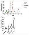
Serum activin A level of patient is independent of BMI as a prognostic tool for AP disease severity. (a) ELISA of serum activin A levels (y axis) plotted against the patient BMI (x axis) distributed by disease severity group (control = inverted triangle; mild AP = square; moderate AP = triangle; severe AP = circle) n = 10 for each group with activin A estimated at admission and 24 hr and 48–72 hr after admission for each patient. (b) Serum activin A levels in those subjects with a BMI ≤30. GEE multivariate analysis confirmed that serum activin A level is predictive of disease severity for patients with low BMI (P < 0.0001). AP, acute pancreatitis; BMI, body mass index; GEE, generalized estimating equation.
Figure 2.
Figure 2.
Inhibition of activin A in cerulein-induced AP murine model reduces disease severity and inflammation. (a) Schematic of treatment and cerulein injection schedule. Mice were pretreated with activin A-specific neutralizing antibody (Anti-Act) or IgG control 30 minutes before the start of cerulein injections. Mice were given 12 hourly injections of 50 μg/kg cerulein or equal volume PBS. Blood and tissue were collected 24 hr after the start of cerulein injections. (b) Stained sections of pancreatic tissue from mice with cerulein-induced AP. Top row, H&E staining; middle row, IHC staining of CD68; and bottom row, IHC staining of TGFβ. Representative images were taken at ×40 total magnification. (c) Quantification of pancreatic tissue staining. n = 5–10 mice per group. *P < 0.05; **P < 0.01; ***P < 0.001. AP, acute pancreatitis; IHC, immunohistochemical; PBS, phosphate-buffered saline.
Figure 3.
Figure 3.
Inhibition of activin A treatment of cerulein-induced AP decreases disease severity but does not reduce pancreas regeneration. (a) Schematic of treatment and cerulein injection schedule. Mice (n = 3–5) were given 12 hourly injections of 50 μg/kg cerulein or equal volume PBS. At 7 hr of cerulein injection, mice were treated with activin A-specific neutralizing antibody (Anti-Act) or IgG control. Blood and tissue were collected 24 hr after the start of cerulein injections and at 7 d. (b) Hematoxylin/eosin stained pancreatic sections at ×10 magnification. (c) Edema score at 24 hr and 7 d of cerulein injection with and without Anti-Act. (d) Percentage of pancreatic necrosis, (e) pancreas:body weight ratio, (f) neutrophil infiltration score, and (g) AP severity score. **P < 0.01, ***P < 0.001. AP, acute pancreatitis; PBS, phosphate-buffered saline.
Figure 4.
Figure 4.
Activin A is produced and released by pancreatic cell lines but not by macrophage cell lines. (a) qRT-PCR results of INHBA mRNA levels in hPSC and HPDE cells after 24-hr treatment with 10 ng/mL TNF. (b) Representative immunoblots of the βA subunit proform of activin A in hPSC and HPDE cells after 24-hr treatment with 10 ng/mL TNF. Loading control is α-tubulin for hPSC cells and β-actin for HPDE cells. (c) Densitometry quantification of immunoblots of at least 3 independent experiments. (d) ELISA of activin A in cell culture media of hPSC and HPDE cells after treatment with increasing concentrations of TNF or LPS for 24 hr. (f) ELISA of activin A in cell culture media of RAW264.7 cells treated with 10 ng/mL TNF or 10 μg/mL LPS for 24 hr. Activin A in the media was undetectable. (e) INHBA mRNA level in RAW264.7 cells after activation with 10 ng/mL TNF. *P < 0.05; **P < 0.01. HPDE, human ductal epithelial cells; hPSC, human pancreas stellate cells; qRT-PCR, quantitative real-time polymerase chain reaction.
Figure 5.
Figure 5.
Activin A activates RAW264.7 macrophages. qRT-PCR results of TNF and IL1B mRNA expression in RAW264.7 macrophages after 24-hour treatment with 25 ng/mL activin A. *P < 0.05. qRT-PCR, quantitative real-time polymerase chain reaction.

References

    1. Peery AF, Crockett SD, Murphy CC, et al. Burden and cost of gastrointestinal, liver, and pancreatic diseases in the United States: Update 2018. Gastroenterology 2019;156(1):254–72.e211. - PMC - PubMed
    1. Yadav D, Lowenfels AB. The epidemiology of pancreatitis and pancreatic cancer. Gastroenterology 2013;144(6):1252–61. - PMC - PubMed
    1. Banks PA, Bollen TL, Dervenis C, et al. Classification of acute pancreatitis—2012: Revision of the Atlanta Classification and Definitions by International Consensus. Gut 2013;62(1):102–11. - PubMed
    1. Beger HG, Rau BM. Severe acute pancreatitis: Clinical course and management. World J Gastroenterol 2007;13(38):5043–51. - PMC - PubMed
    1. Garg PK, Madan K, Pande GK, et al. Association of extent and infection of pancreatic necrosis with organ failure and death in acute necrotizing pancreatitis. Clin Gastroenterol Hepatol 2005;3(2):159–66. - PubMed

Publication types

MeSH terms