Review
. 2020 Nov;77(22):4459-4483.
doi: 10.1007/s00018-020-03536-5. Epub 2020 May 1.

The multifaceted role of reactive oxygen species in tumorigenesis

Affiliations
Review

The multifaceted role of reactive oxygen species in tumorigenesis

Anuradha Kirtonia et al. Cell Mol Life Sci. 2020 Nov.

Abstract

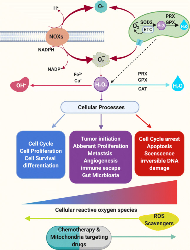
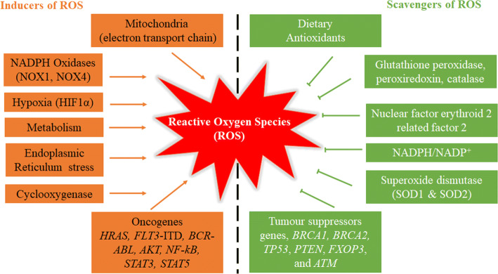
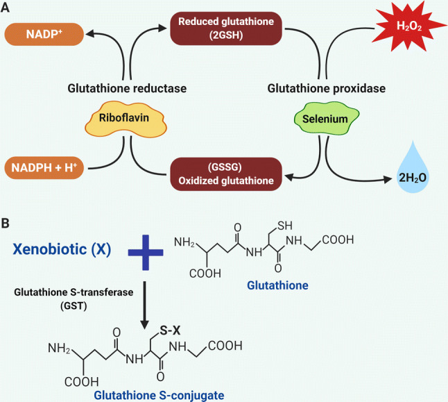
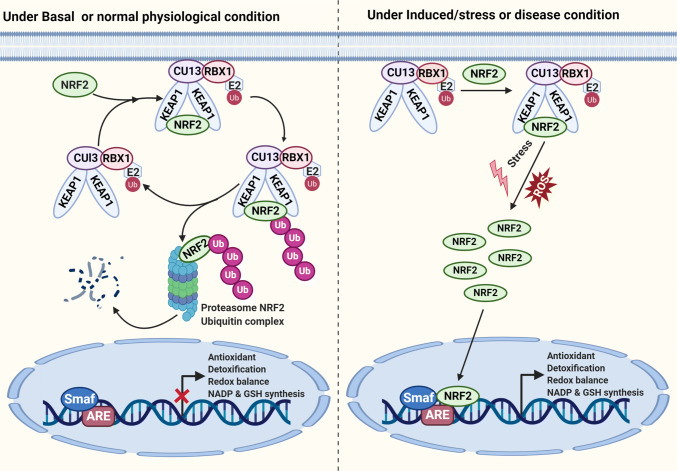
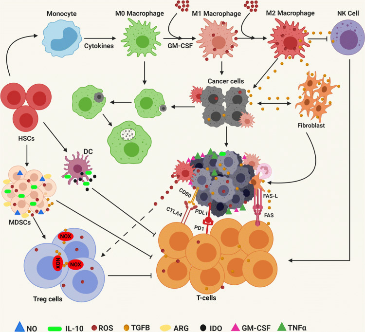
Redox homeostasis is an essential requirement of the biological systems for performing various normal cellular functions including cellular growth, differentiation, senescence, survival and aging in humans. The changes in the basal levels of reactive oxygen species (ROS) are detrimental to cells and often lead to several disease conditions including cardiovascular, neurological, diabetes and cancer. During the last two decades, substantial research has been done which clearly suggests that ROS are essential for the initiation, progression, angiogenesis as well as metastasis of cancer in several ways. During the last two decades, the potential of dysregulated ROS to enhance tumor formation through the activation of various oncogenic signaling pathways, DNA mutations, immune escape, tumor microenvironment, metastasis, angiogenesis and extension of telomere has been discovered. At present, surgery followed by chemotherapy and/or radiotherapy is the major therapeutic modality for treating patients with either early or advanced stages of cancer. However, the majority of patients relapse or did not respond to initial treatment. One of the reasons for recurrence/relapse is the altered levels of ROS in tumor cells as well as in cancer-initiating stem cells. One of the critical issues is targeting the intracellular/extracellular ROS for significant antitumor response and relapse-free survival. Indeed, a large number of FDA-approved anticancer drugs are efficient to eliminate cancer cells and drug resistance by increasing ROS production. Thus, the modulation of oxidative stress response might represent a potential approach to eradicate cancer in combination with FDA-approved chemotherapies, radiotherapies as well as immunotherapies.

Keywords: Angiogenesis; Antioxidant system; Cancer stem cells (CSCs); Chemotherapy; Ferroptosis; Immune escape; Metastasis; Mitochondrial ROS (mROS); ROS scavenger; Reactive oxygen species (ROS); Signaling pathways; Tumor microenvironment.


Conflict of interest statement

All the authors have read the manuscript and have no competing interests.

Figures

Fig. 1
Fig. 1
Formation and regulation of ROS and its effects on cellular functions. Mitochondria and NADPH oxidases are major sources of O2, HO•, and H2O2 (ROS) formation. Superoxide dismutase (SOD1 or SOD2) can convert O2 into H2O2. H2O2 can be converted into H2O (water) by peroxiredoxin (PRX), glutathione peroxidase (GPX) and catalase (CAT) in mitochondria and cytosol. ROS are generated during normal cellular functioning and homeostasis is maintained by antioxidants expressed by the cells. Low ROS (green) is the basic need to maintain normal cellular proliferation, survival, and differentiation. Moderate to high ROS (tumor favoring ROS; light red) is the signal for the increased cellular proliferation, survival, tumor initiation, immune escape to genomic instability, metastasis, invasion and angiogenesis. Extremely high ROS produced by chemotherapeutic agents (dark red) is dangerous for the cells and leads to cell cycle arrest, apoptosis, senescence and unrepairable DNA damage
Fig. 2
Fig. 2
Maintenance of cellular homeostasis through inducers and scavengers of ROS. ROS can be produced by mitochondria, NADPH oxidases, hypoxia, metabolism, ER stress, cyclooxygenase and oncogenes including HRAS, FLT3-ITD, BCR-ABL, AKT, NF-kB, STAT3 and STAT5. On the other hand, ROS can be eliminated via activation of the dietary antioxidants, glutathione peroxidase, peroxiredoxin, catalase, NRF2, NADPH, SOD and tumor suppressor gnes including BRCA1, BRCA2, TP53, PTEN, FXOP3 and ATM
Fig. 3
Fig. 3
Glutathione antioxidant system. a Schematics for the reduction of hydrogen peroxide. Nicotinamide Adenine Dinucleotide Phosphate is essential for the regeneration of GSH via glutathione reductase. Hydrogen peroxide (H2O2) is reduced to water (H2O) via glutathione peroxidase. b Mechanism of the glutathione S transferases (GSTs). Glutathione conjugation with xenobiotic (X) is mainly catalyzed via GST to from glutathione S conjugate
Fig. 4
Fig. 4
Role of the NRF2/KEAP1 antioxidant pathway for maintaining cellular homeostasis. Under normal physiological condition, NRF2 interact with KEAP1 to activate Cul3‐dependent ubiquitination and its degradation via the proteasome. Under stress or induced condition, NRF2 dissociates from KEAP1 and translocates into the nucleus. NRF2 forms a heterodimer with sMaf protein as well as to ARE to initiate the transcription of several downstream genes
Fig. 5
Fig. 5
Mechanism and inducer of ferroptosis. Suppression of system Xc/GPX4 activity caused ferroptosis to induce cell death. Elevation of lipid ROS results in the ferroptopsis
Fig. 6
Fig. 6
ROS activate RAS and PI3/AKT signaling pathways. Growth factor receptor signaling can generate ROS through growth factors, NOXs and mitochondria. ROS can activate RAS/MAPK and PI3K/AKT/mTOR signaling cascade either though inactivation of phosphatases such as PTEN or PTP at cysteine residues or by direct oxidation of kinases. Other mechanisms by which ROS induce cellular signaling are through activation NF-kB signaling
Fig. 7
Fig. 7
ROS-dependent STAT3 pathway in metastasis and drug resistance. Growth factors, ionizing radiation, mitochondria and NOX4 result in the production of intracellular ROS. ROS activate cancer cells and cancer-associated fibroblast cells to secrete IL-6. IL-6 activates the STAT3 pathway and promotes tumor metastasis, resistance to chemotherapy and radiotherapy, and CSC self-renewal
Fig. 8
Fig. 8
Mitochondrial ROS in hypoxia and angiogenesis. In oxygen-rich conditions, HIF-1α forms complex with VHL with the help of PHD2. This results in ubiquitination and proteasome-mediated degradation of the complex. On the other hand, mROS can cause the depletion of oxygen levels and inhibition of PHD2 activity resulting in HIF-1 α stabilization, by forming a dimer with HIF-1β. This dimer moves to the nucleus and results in transcriptional activation of VEGF, EPO
Fig. 9
Fig. 9
Involvement of ROS in tumor microenvironment and immunosuppression. Myeloid-derived suppressor cells (MDSCs) are generated due to secretion of growth factors (GM-CSF, M-CSF, VEGF) and pro-inflammatory cytokines (IFN-ϒ, IL-1β, IL-4, TNFα by tumor cells. MDSC secrete ROS, nitric oxide (NO) and arginase (ARG) to inactivate T cell and TGFβ, and IL10 to activate regulatory T cells (Tregs). ROS convert M0 macrophages into TAMs and secrete immune-suppressive factors and cytokines to block NK and CTLs. Tumor cells and stromal cells express TGFβ, checkpoint ligands and FasL to cause T cell apoptosis. ROS help tumor cells to overexpress PDL1/2 and CTLA4 to inhibit CTLs. TGFβ stimulates NOXs within the Treg cells to trigger ROS production. Macrophage-induced ROS leads to the accumulation of Treg cells. MDSC produces a large amount of ROS to trigger Tregs and suppress T cells

References

    1. Gorrini C, Harris IS, Mak TW. Modulation of oxidative stress as an anticancer strategy. Nat Rev Drug Discov. 2013;12:931–947. - PubMed
    1. D'Autreaux B, Toledano MB. ROS as signalling molecules: mechanisms that generate specificity in ROS homeostasis. Nat Rev Mol Cell Biol. 2007;8:813–824. - PubMed
    1. Handy DE, Loscalzo J. Redox regulation of mitochondrial function. Antioxid Redox Signal. 2012;16:1323–1367. - PMC - PubMed
    1. Cross CE, Halliwell B, Borish ET, Pryor WA, Ames BN, Saul RL, McCord JM, Harman D. Oxygen radicals and human disease. Ann Intern Med. 1987;107:526–545. - PubMed
    1. Brewer TF, Garcia FJ, Onak CS, Carroll KS, Chang CJ. Chemical approaches to discovery and study of sources and targets of hydrogen peroxide redox signaling through NADPH oxidase proteins. Annu Rev Biochem. 2015;84:765–790. - PMC - PubMed

Substances