doi: 10.1016/S1473-3099(20)30367-4.
Epub 2020 Apr 30.
Hypoxaemia related to COVID-19: vascular and perfusion abnormalities on dual-energy CT
Affiliations
- PMID: 32359410
- PMCID: PMC7252023
- DOI: 10.1016/S1473-3099(20)30367-4
Item in Clipboard
Hypoxaemia related to COVID-19: vascular and perfusion abnormalities on dual-energy CT
Lancet Infect Dis.
2020 Dec.
No abstract available
Figures
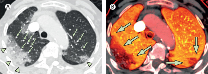
Dual-energy CT in a patient with COVID-19 pneumonia without evidence of pulmonary emboli Patient 1, an 87-year-old woman with a history of fever and cough for 5 days, was found on the floor of her nursing home. On admission to hospital, the patient required a non-rebreather mask with a flow rate of 15 L/min to maintain an oxygen saturation of 85%; intubation was not pursued as the patient's status was comfort measures only. (A) There is a large area of peripheral ground-glass opacity and consolidation within the right upper lobe and smaller ground-glass opacity in the posterior left upper lobe (green arrowheads), which are accompanied by dilated subsegmental vessels proximal to, and within, the opacities (green arrows). (B) The accompanying image of pulmonary blood volume shows corresponding wedge-shaped areas of decreased perfusion within the upper lobes, with a peripheral halo of higher perfusion (green arrows). COVID-19=coronavirus disease 2019.
Comment in
-
Reversibility of venous dilatation and parenchymal changes density in Sars-Cov-2 pneumonia: toward the definition of a peculiar pattern.Pulmonology. 2021 Jul-Aug;27(4):353-357. doi: 10.1016/j.pulmoe.2020.10.010. Epub 2020 Nov 16. Pulmonology. 2021. PMID: 33272912 Free PMC article. No abstract available.
References
-
- Otrakji A, Digumarthy SR, Lo Gullo R, Flores EJ, Shepard JA, Kalra MK. Dual-energy CT: spectrum of thoracic abnormalities. Radiographics. 2016;36:38–52. - PubMed
Publication types
MeSH terms
Substances
LinkOut - more resources
Full Text Sources
Other Literature Sources
Medical