. 2020 Jun 8;30(11):2175-2183.e6.
doi: 10.1016/j.cub.2020.03.070. Epub 2020 Apr 30.

CLASP Mediates Microtubule Repair by Restricting Lattice Damage and Regulating Tubulin Incorporation

Affiliations

CLASP Mediates Microtubule Repair by Restricting Lattice Damage and Regulating Tubulin Incorporation

Amol Aher et al. Curr Biol. .

Abstract

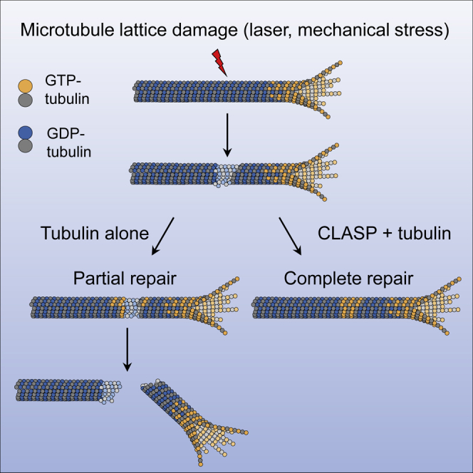
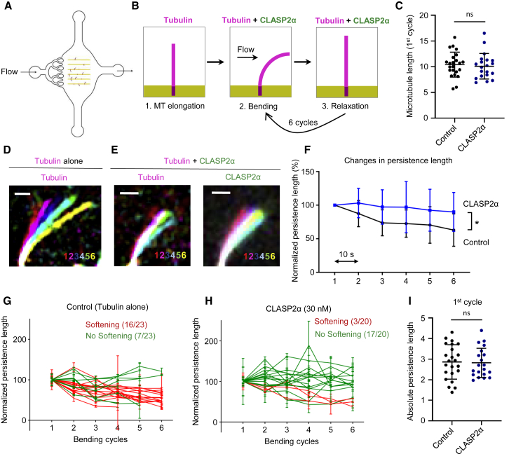
Microtubules play a key role in cell division, motility, and intracellular trafficking. Microtubule lattices are generally regarded as stable structures that undergo turnover through dynamic instability of their ends [1]. However, recent evidence suggests that microtubules also exchange tubulin dimers at the sites of lattice defects, which can be induced by mechanical stress, severing enzymes, or occur spontaneously during polymerization [2-6]. Tubulin incorporation can restore microtubule integrity; moreover, "islands" of freshly incorporated GTP-tubulin can inhibit microtubule disassembly and promote rescues [3, 4, 6-8]. Microtubule repair occurs in vitro in the presence of tubulin alone [2-6, 9]. However, in cells, it is likely to be regulated by specific factors, the nature of which is currently unknown. CLASPs are interesting candidates for microtubule repair because they induce microtubule nucleation, stimulate rescue, and suppress catastrophes by stabilizing incomplete growing plus ends with lagging protofilaments and promoting their conversion into complete ones [10-17]. Here, we used in vitro reconstitution assays combined with laser microsurgery and microfluidics to show that CLASP2α indeed stimulates microtubule lattice repair. CLASP2α promoted tubulin incorporation into damaged lattice sites, thereby restoring microtubule integrity. Furthermore, it induced the formation of complete tubes from partial protofilament assemblies and inhibited microtubule softening caused by hydrodynamic-flow-induced bending. The catastrophe-suppressing domain of CLASP2α, TOG2, combined with a microtubule-tethering region, was sufficient to stimulate microtubule repair, suggesting that catastrophe suppression and lattice repair are mechanistically similar. Our results suggest that the cellular machinery controlling microtubule nucleation and growth can also help to maintain microtubule integrity.

Keywords: CLASP; TOG2; in vitro reconstitution; laser microsurgery; lattice defects; microtubule catastrophe; microtubule dynamics; microtubule repair; microtubule rescue; tubulin.


Conflict of interest statement

Declaration of Interests The authors declare no competing interests.

Figures

None
Graphical abstract
Figure 1
Figure 1
CLASP Promotes Repair of Microtubule Lattices Damaged by Laser Illumination (A) A scheme of full-length CLASP2α and its TOG2-S fragment. Vertical lines labeled SxIP (Ser-any amino acid-Ile-Pro) represent EB-binding motifs located in the unstructured positively charged region adjacent to the TOG2 domain. (B) Schematic for an experiment to monitor the possible outcomes of a 532 nm pulsed laser induced damage at a site along the dynamic lattice. (I) Microtubule bending at the site of damage, which can lead to either straightening of the lattice or microtubule breakage. (II) Reduction of the tubulin intensity. (III) Microtubule severing resulting in direct appearance of two microtubule ends. (C) Stills from a time-lapse video showing photodamage of a dynamic microtubule grown in the presence of Rhodamine-tubulin alone followed by bending and subsequent breakage (outcome I). Scale bar, 2 μm. (D) Percentage of total events for outcome I resulting in either microtubule breakage or straightening at the point of photodamage in the presence of tubulin alone (n = 22 microtubules analyzed from 4 experiments) or together with either 30 nM GFP-CLASP2α (n = 53 microtubule analyzed from 6 experiments) or 30 nM GFP-TOG2-S (n = 54 from 8 experiments). Error bars denote SD. (E and F) Normalized mean intensity at the site of photodamage in case of outcome I for the GFP channel for CLASP2α and TOG2-S (E) and Rhodamine-tubulin channel in the presence of tubulin alone or together with either CLASP2α or TOG2-S (F); before damage (black), immediately after damage (orange) and after microtubule straightening (blue). Tubulin alone: n = 4 microtubules, 4 experiments; CLASP2α: n = 21 microtubules, 4 experiments; TOG2-S: n = 20 microtubules, 6 experiments. Error bars denote SD. (G) Stills from a time-lapse video showing a dynamic microtubule grown in the presence of Rhodamine-tubulin together with 30 nM GFP-CLASP2α for outcome I. Normalized intensity profiles along the microtubule for the CLASP (green) and tubulin channel (magenta) at different time points are shown in the bottom panels, with the arrow pointing to the site of photodamage. The purple circle on the plot indicates the end of the microtubule. Scale bars, 2 μm. (H) Normalized mean tubulin fluorescence intensity over time at the site of local photodamage (outcome II); microtubules were grown in the presence of Rhodamine tubulin alone (gray) (n = 35 microtubules, 2 experiments) or together with 30 nM GFP-CLASP2α (blue) (n = 44 microtubules, 2 experiments). Straight lines were fitted to the initial increase in tubulin intensity until saturation for the respective mean values yielding slopes as indicated. (I and J) Stills and the corresponding kymograph of a microtubule grown in the presence of Rhodamine-tubulin alone (I) or together with GFP-CLASP2α (30 nM) (J) severed with a 532 nm laser as indicated (outcome III). Scale bars: still image, 2 μm; kymograph, 4 μm (horizontal) and 10 s (vertical). Dotted yellow lines point to the time point of the still in the kymograph. (K) Percentage of total laser severing events resulting in either immediate microtubule regrowth at the site of photoablation, microtubule depolymerization to the seed or depolymerization followed by rescue along the lattice, in the presence of Rhodamine-tubulin alone or together with either 30 nM GFP-CLASP2α or 30 nM GFP-TOG2-S. Tubulin alone: n = 186 microtubules, 3 experiments: CLASP2α: n = 36 microtubules, 3 experiments; TOG2-S: n = 48 microtubules, 8 experiments. For plots in Figure 1D: p = 0.0091, ∗∗p = 0.0381, for Figures 1E and 1F, ∗∗∗∗p < 0.0001, ∗∗∗p = 0.001 Mann-Whitney U test. See also Figures S1 and S2, Table S1, and Videos S1 and S2.
Figure 2
Figure 2
CLASP Promotes Formation of Complete Microtubules from Partial Protofilament Assemblies (A) Cartoon illustrating the changes in tubulin sheet- or ribbon-like structures generated at the plus ends of dynamic microtubules in the presence of 30 nM kinesin-5-GFP dimer (Kin-5 dimer) alone or together with 30 nM GFP-CLASP2α. (B) Stills from a time-lapse video showing a plus end of a microtubule grown in the presence of Rhodamine-tubulin and 30 nM Kin-5-GFP dimer. Scale bar: 2 μm. (C) Mean tubulin intensity values for the straight and the curled portions of the microtubule lattice as indicated for microtubules grown in the presence of Rhodamine tubulin and 30 nM Kin-5-GFP dimer. n = 25 microtubules from 2 experiments. Error bars denote SD. (D) Number of newly generated microtubule ends from a single microtubule plus end for microtubules grown in the presence of Rhodamine-tubulin and 30 nM Kin-5-GFP dimer alone (n = 38 microtubule plus ends, 3 experiments) or together with 30 nM TagBFP-CLASP2α (n = 95 microtubule plus ends, 3 experiments), or with 30 nM TagBFP-TOG2-S (n = 85 microtubule plus ends, 3 experiments), or with 100 nM Tag-BFP-TOG2-S (n = 26 microtubule plus ends, 2 experiments). Events where the microtubule plus ends bent by angles over 45° with respect to the lattice were monitored in a 10 min time lapse. Error bars denote SD. (E) Stills from a time-lapse video showing the plus end for a microtubule grown in the presence of Rhodamine tubulin, 30 nM Kin-5-GFP dimer, and 30 nM TagBFP-CLASP2α. Scale bar: 2 μm. (F) Stills from a time-lapse video showing a microtubule plus end grown in the presence of Rhodamine-tubulin, 30 nM Kin-5-GFP dimer together with 30 nM TagBFP-TOG2-S. Scale bar: 3 μm. For (B), (E), and (F), green arrowheads point to the plus end and blue arrowheads to the minus ends. For all plots, ∗∗∗∗p < 0.0001, ∗∗∗p = 0.001 and ns, no significant difference with control, Mann-Whitney U test. See also Video S3.
Figure 3
Figure 3
CLASP Promotes Complete Repair of Damaged Microtubule Lattices (A) Schematic for an experiment to monitor tubulin incorporation into damaged microtubule lattices. Microtubules prepared from Rhodamine-tubulin in the presence of Taxol were first incubated without Taxol and tubulin for 1.5 min and subsequently with 5 μM HiLyte Fluor-488-labeled tubulin with or without 30 nM mCherry-CLASP2α or 30 nM mCherry-TOG2-S. After 10 min, the residual free green tubulin was washed out with the wash buffer supplemented with 25% glycerol to prevent microtubule depolymerization, in order to better visualize incorporation of green tubulin. (B–D) Microtubule repair in the presence of tubulin alone (B) or together with either 30 nM mCherry-CLASP2α (C) or 30 nM mCherry-TOG2-S (D). Single frames (top) of time-lapse videos after the final washout and kymographs (bottom) showing green tubulin incorporation sites (numbered asterisks in stills) into Rhodamine-labeled microtubule lattices (magenta). In kymographs, white arrows indicate complete repair and white arrowheads partial repair. Enlarged views of the boxed regions in the kymographs in (B)–(D) showing partial (B) or complete microtubule repair (C and D) in the bottom-left panel for each condition. Yellow arrowheads in (B) (bottom left) indicate events of loss of freshly incorporated tubulin. Intensity profiles along the microtubule for the Rhodamine-labeled microtubule seed channel (magenta) with or without mCherry-CLASP2α, and for the green tubulin channel are shown in the bottom-right panel for each condition. The numbers indicate incorporation sites specified in stills in (B) and (D). Scale bars: 2 μm (horizontal) and 60 s (vertical). See also Video S4. (E and G) Frequency of incorporation per unit length per microtubule (E) and the average length of incorporations (G) in the presence of tubulin alone (n = 73, M = 25, L = 459.25 μm, 2 experiments), together with 30 nM mCherry-CLASP2α (n = 64, M = 31, L = 450.74 μm, 5 experiments) or 30 nM mCherry-TOG2-S (n = 52, M = 37, L = 418.43 μm, 4 experiments), where n, M, and L are total number of incorporations, total number of microtubules, and total length of microtubules analyzed, respectively. Error bars represent SEM. ∗∗p = 0.0038 and ns, no significant difference with control, Mann-Whitney U test. (F) Fraction of total events resulting in either complete or partial repair at the site of tubulin incorporations with the length exceeding 1 μm, in the presence of tubulin alone (n = 15, 2 experiments), together with either 30 nM mCherry-CLASP2α (n = 22, 5 experiments) or 30 nM mCherry-TOG2-S (n = 23, 4 experiments), where n is the total number of incorporations longer than 1 μm. See also Video S4.
Figure 4
Figure 4
CLASP2α Inhibits Microtubule Softening Induced by Hydrodynamic Flow (A) Illustration of the microfluidic device used for microtubule bending. (B) Scheme of the work sequence: 1. Red fluorescent microtubules were grown from seeds grafted on micropatterned lines. 2. Microtubules were bent for 10 s by applying a fluid flow using the same mix as in 1, with or without 30 nM GFP-CLASP2α. 3. The flow was stopped for 10 s before repeating the bending cycle. (C) Lengths of microtubules used in the bending experiments in the presence of tubulin alone (n = 23 microtubules) or together with 30 nM GFP-CLASP2α (n = 20 microtubules). Error bars denote SD. (D and E) Images showing an overlay of maximum bent conformations where every cycle is represented in a different color for the tubulin channel for a microtubule bent in the presence of tubulin alone (D) and for both tubulin and CLASP channels for a microtubule bent in the presence of tubulin together with 30 nM GFP-CLASP2α (E). Scale bar: 3 μm. (F) Persistence length measured for microtubules bent in the presence (blue curve, n = 20) or absence (black curve, n = 23) of 30 nM GFP-CLASP2α. Persistence length was normalized to the value in the first bending cycle for each microtubule. Values represent the average persistence length (mean ± SD) of individual measurements shown in Figures 4G and 4H. To test whether microtubules showed softening, a Spearman correlation test for persistence length values over subsequent bending cycles was performed. It revealed significant softening of microtubules in both conditions (p = 0.01 and 0.08, respectively), though it was much less pronounced in the presence of CLASP2α. A t test confirmed significant difference between the two curves (p = 0.006). (G and H) Individual persistence length measurements of microtubules bent in the absence (G) and presence (H) of 30 nM GFP-CLASP2α. Values represent the average of five independent measurements for each bending cycle (mean ± SD) and were normalized to the initial value. A Spearman correlation test was performed to test for softening. Red lines indicate microtubules that became significantly softer, and green lines indicate microtubules that did not show significant softening. (I) Absolute persistence lengths for microtubules bent in the presence of tubulin alone (n = 23 microtubules) or together with 30 nM GFP-CLASP2α (n = 20 microtubules) after the 1st cycle. Error bars denote SD. See also Video S5.

Comment in

References

    1. Desai A., Mitchison T.J. Microtubule polymerization dynamics. Annu. Rev. Cell Dev. Biol. 1997;13:83–117. - PubMed
    1. Schaedel L., John K., Gaillard J., Nachury M.V., Blanchoin L., Théry M. Microtubules self-repair in response to mechanical stress. Nat. Mater. 2015;14:1156–1163. - PMC - PubMed
    1. Aumeier C., Schaedel L., Gaillard J., John K., Blanchoin L., Théry M. Self-repair promotes microtubule rescue. Nat. Cell Biol. 2016;18:1054–1064. - PMC - PubMed
    1. Schaedel L., Triclin S., Chrétien D., Abrieu A., Aumeier C., Gaillard J., Blanchoin L., Théry M., John K. Lattice defects induce microtubule self-renewal. Nat. Phys. 2019;15:830–838. - PMC - PubMed
    1. Aher A., Akhmanova A. Tipping microtubule dynamics, one protofilament at a time. Curr. Opin. Cell Biol. 2018;50:86–93. - PubMed

Publication types

LinkOut - more resources