. 2020 Aug;38(8):947-953.
doi: 10.1038/s41587-020-0462-y. Epub 2020 Mar 23.

Human chimeric antigen receptor macrophages for cancer immunotherapy

Affiliations

Human chimeric antigen receptor macrophages for cancer immunotherapy

Michael Klichinsky et al. Nat Biotechnol. 2020 Aug.

Abstract

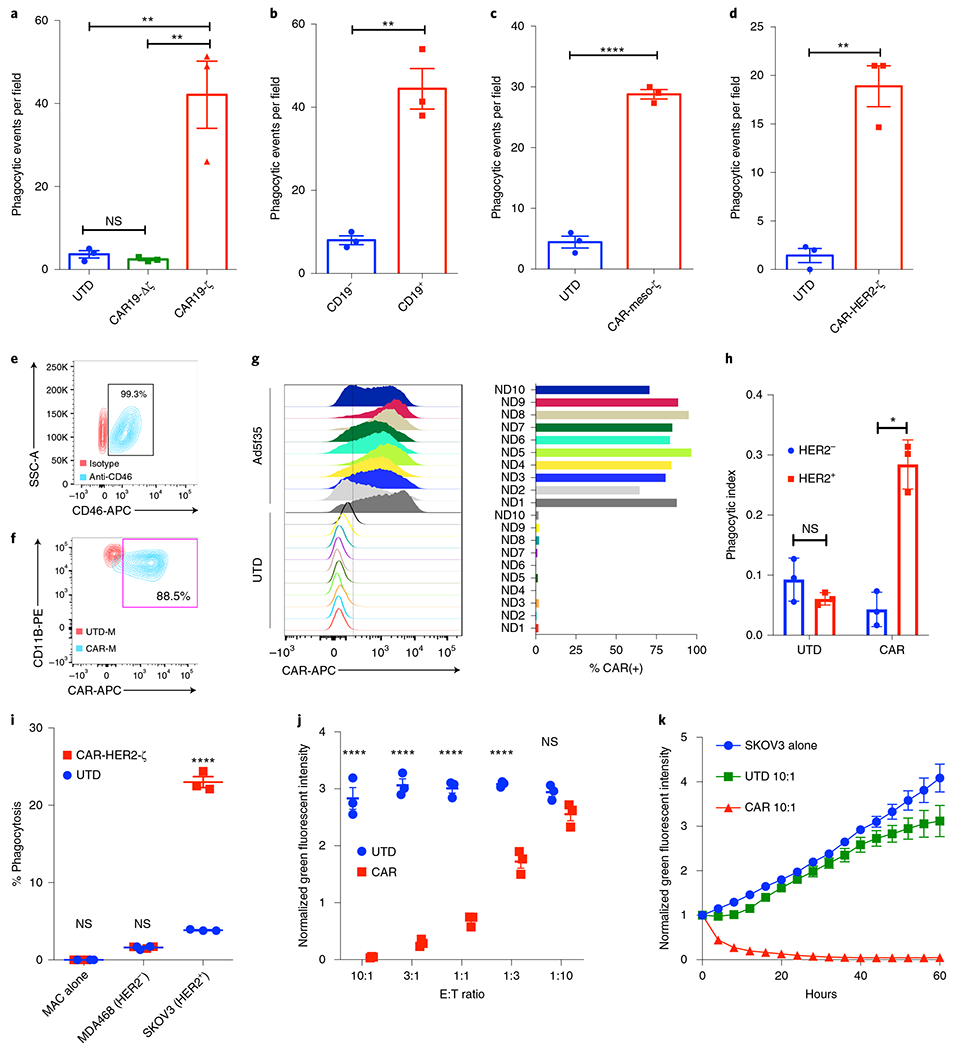
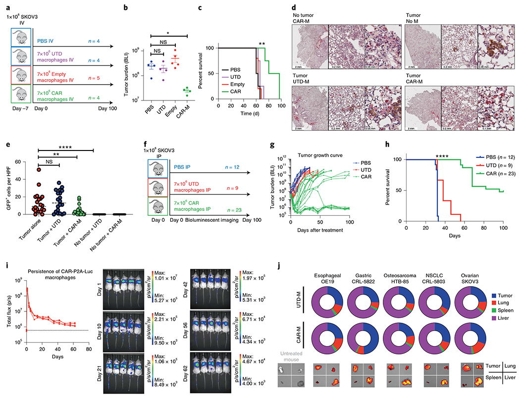
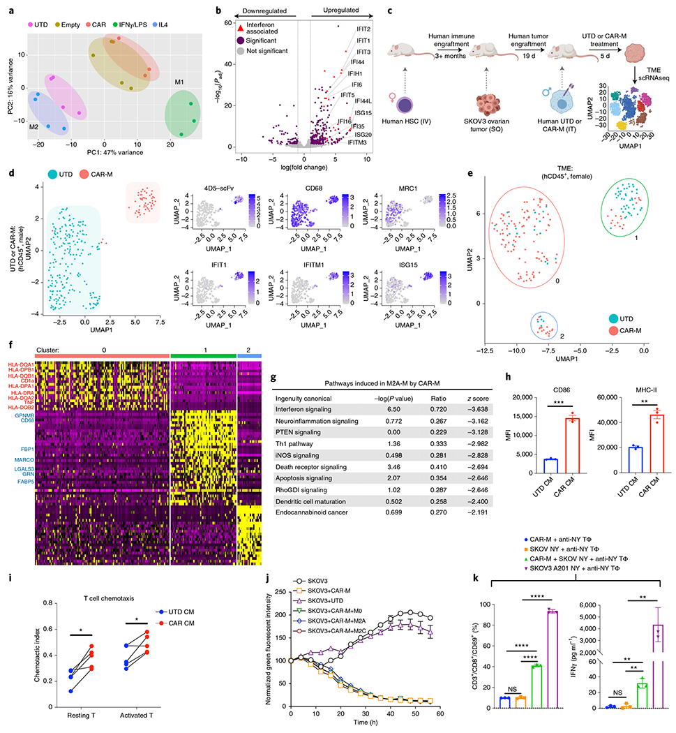
Chimeric antigen receptor (CAR) T cell therapy has shown promise in hematologic malignancies, but its application to solid tumors has been challenging1-4. Given the unique effector functions of macrophages and their capacity to penetrate tumors5, we genetically engineered human macrophages with CARs to direct their phagocytic activity against tumors. We found that a chimeric adenoviral vector overcame the inherent resistance of primary human macrophages to genetic manipulation and imparted a sustained pro-inflammatory (M1) phenotype. CAR macrophages (CAR-Ms) demonstrated antigen-specific phagocytosis and tumor clearance in vitro. In two solid tumor xenograft mouse models, a single infusion of human CAR-Ms decreased tumor burden and prolonged overall survival. Characterization of CAR-M activity showed that CAR-Ms expressed pro-inflammatory cytokines and chemokines, converted bystander M2 macrophages to M1, upregulated antigen presentation machinery, recruited and presented antigen to T cells and resisted the effects of immunosuppressive cytokines. In humanized mouse models, CAR-Ms were further shown to induce a pro-inflammatory tumor microenvironment and boost anti-tumor T cell activity.


Conflict of interest statement

Competing interests

M.K. and S.G. are scientific co-founders and Scientific Advisory Board members and hold equity in Carisma Therapeutics. C.J. is a member of the Scientific Advisory Board of Carisma Therapeutics. M.K., S.G. and C.J. are inventors on intellectual property related to this work. S.G. has received research funding from Carisma Therapeutics. A.B., M.Z., M.S., K.G., N.A., Y.O. and M.K. are employees of Carisma Therapeutics. Carisma Therapeutics is a company pursuing the commercial development of this technology.

Figures

Fig. 1 |
Fig. 1 |. Generation of human CAR-Ms and assessment of CAR-mediated tumor phagocytosis.
a, Assessment of indicated THP-1 macrophage phagocytosis against CD19+ K562 target cells using fluorescent microscopy. Data are represented as the mean ± s.e.m. of n = 3 technical triplicates, with three random fields of view assessed per replicate. Statistical significance was calculated with one-way ANOVA with multiple comparisons. b, Phagocytosis of CD19+ or control CD19 K562 target cells by CAR19ζ-expressing THP-1 macrophages. Data are represented as the mean ± s.e.m. of n = 3 technical triplicates, with three random fields of view assessed per replicate. Statistical significance was calculated using a two-sided t-test. c,d, In vitro phagocytosis by UTD or CAR-meso-ζ THP-1 macrophages of mesothelin+ve K562 cells (c) or by CAR-HER2-ζ macrophages of HER2+ K562 cells (d). Data are represented as mean ± s.e.m. of n = 3 technical replicates, with three random fields of view assessed per replicate. Statistical significance was calculated using a two-sided t-test. e, Representative FACS plot of CD46 expression on primary human macrophages (blue) or isotype control (red). This graph is representative of n = 10 human donors. f, Representative FACS plot of CAR-HER2 expression on Ad5f35-transduced primary human macrophages (blue, CAR-M; red, UTD-M). This graph is representative of n = 10 human donors. g, CAR expression on macrophages derived from n = 10 human donors 48 h after Ad5f35-CAR transduction. h, Incucyte-based phagocytosis of HER2 functionalized or control pH-Rodo-labeled beads by UTD or CAR-M. Data are represented as mean ± s.e.m. of n = 3 technical replicates and are representative of three experiments. Statistical analysis was calculated using a two-sided t-test. i, FACS-based phagocytosis of HER2 (MDA-468) or HER2+ (SKOV3) target tumor cells by UTD or CAR-M. Statistical significance was calculated with one-way ANOVA with multiple comparisons, and data represent n = 3 technical replicates (representative of at least three individual experiments). j, Incucyte-based killing assay of GFP+ SKOV3 by UTD or CAR-M after 48 h of co-culture at different E:T ratios. Statistical significance was calculated with one-way ANOVA with multiple comparisons, and data represent the mean ± s.e.m. of n = 3 technical replicates (representative of at least three individual experiments). k, Incucyte-based killing kinetics of UTD or CAR-M against SKOV3 at a 10:1 E:T ratio. Data are represented as the mean ± s.e.m. of n = 3 technical replicates (representative of at least three individual experiments). For all panels, *P < 0.05, **P < 0.01, ***P < 0.001, ****P < 0.0001.
Fig. 2 |
Fig. 2 |. Evaluation of anti-tumor activity, persistence and trafficking of primary human anti-HER2 CAR-Ms in xenograft models.
a, NSGS mice were IV injected with luciferase(+) SKOV3 and treated with IV PBS, UTD, empty vector transduced (empty) or CAR-HER2 macrophages 7 d later with n as shown. b, Tumor burden (total flux) by bioluminescent imaging 31 d after treatment. Data are represented as mean ± s.e.m., and statistical significance was calculated with ANOVA with multiple comparisons. c, Kaplan-Meier survival curve (median survival, 88.5 d (CAR) versus 63 d (Empty), P = 0.0047). Statistical significance was calculated using the log-rank Mantel-Cox test with df = 2. d, Lungs from mice with metastatic disease were assessed with dual immunohistochemical analysis (tumor: anti-GFP, brown; macrophage detection: anti-human CD68, red). Each panel shows low (left), medium (middle) and high (right) power view. The experiment was performed once. e, Quantification of GFP+ tumor cells in n = 5 random ×20 high-power fields per slice, with two slices per mouse and two mice per group. Analysis was done by an observer who was blinded to treatment group allocation. Data are presented as the mean, with statistical significance calculated using multiple two-sided t-tests. f, NSGS mice were injected with SKOV3 IP 2-4 h before receiving injections of PBS, control (UTD) or CAR-HER2 human macrophages IP as shown. g, Tumor burden measured by bioluminescence (total flux) over 100 d. h, Kaplan-Meier survival curve over 100 d. Statistical significance was calculated using the log-rank Mantel-Cox test (CAR versus UTD, P < 0.0001 with df = 2). i, Persistence and biodistribution of CAR-P2A-luciferase-expressing human macrophages after IV administration in n = 5 tumor-free NSGS mice. Luciferase signal was tracked over the course of 62 d (dotted line represents background luminescence). Macrophages initially transited through the lung and accumulated in the liver of tumor-free mice. The experiment was performed twice with similar results. j, Tumor trafficking and biodistribution of VivoTrack680-labeled UTD or CAR-M in five tumor xenograft models. NSGS mice were killed and organs/tumors were explanted for ex vivo imaging 5 d after a single injection of 5 × 10⁶ UTD or CAR-M, n = 3–5 mice per treatment group per tumor type; the experiment was performed once. For all panels, *P < 0.05, **P < 0.01, ***P < 0.001, ****P < 0.0001.
Fig. 3 |
Fig. 3 |. Adenovirally transduced CAR-Ms are M1 polarized, modify the TME and cross-present tumor antigens.
a, Gene expression principal component analysis clustering from UTD, Ad5f35-empty-vector transduced, Ad5f35-CAR-HER2-ζ transduced, classically activated M1 or alternatively activated M2 human macrophages. n = 3–4 human donors. b, Volcano plot of differentially expressed genes in UTD versus CAR-M. Purple indicates Padj < 0.05 and log2 fold change >1 or <−1 Red triangles indicate significantly upregulated interferon-associated genes (n = 4 human donors). Statistical significance was calculated using the Wald test for DESeq2 data. c, Schema of HIS TME model generation. Mice were engrafted with human female CD34+ cells, and after human myeloid engraftment was confirmed, SKOV3 tumors were established in the subcutaneous flank. Nineteen days after tumor administration, 1 × 10⁷ UTD or CAR macrophages from a male human donor were injected intratumorally. Tumors were harvested 5 d after treatment, and scRNAseq was performed on the tumor samples. Adoptively transferred macrophages were distinguished from macrophages in the TME by Y-chromosome gene mapping. d, Cluster plots demonstrating the phenotypic distinction of UTD from CAR macrophages within the TME (left). On the right, the relative expression of the indicated genes was mapped onto CAR-M (top cluster) or UTD (bottom cluster). The data represent three mice and two mice for the CAR-M and UTD arms, respectively. e, Cluster plots demonstrating the phenotypic distinction of human TME after UTD versus CAR-M treatment. Cluster 0 was enriched in the CAR-M-treated arm (86% events from the CAR cohort), whereas cluster 1 was enriched in events from the UTD-treated arm (71% events from the UTD cohort). Cluster 2 was mixed and had few events. f, Gene expression heat map from each TME scRNAseq cluster from e. Pro-inflammatory genes were found in cluster 0 (annotated in red). Potential anti-inflammatory or M2-associated genes in cluster 1 are annotated in blue. For panels c-f, n = 3 mice per group; the experiment was performed once. g, Ingenuity pathway analysis of the transcriptome of in vitro-polarized M2A macrophages after exposure to conditioned media from UTD or CAR-M for 48 h; n = 3 technical replicates. Statistical significance was calculated using Fisher’s exact test. h, Human immature dendritic cells were treated with UTD or CAR-M conditioned media for 48 h, and activation markers CD86 and MHC-II (HLA-DR) were assessed by FACS. Data are represented as the mean ± s.e.m. from n = 3 technical replicates. Statistical significance was calculated using a two-tailed t-test. i, Chemotaxis of resting or activated CD3+ T cells by UTD or CAR-M conditioned media. Data represent T cells from n = 4 human donors, and statistical significance was calculated using a paired t-test. j, Incucyte-based killing assay of SKOV3 by CAR-M alone or in the presence of M0, M2a or M2c macrophages at an E:T:M2 ratio of 3:1:1. Data are represented as the mean ± s.e.m. of n = 3 technical replicates. k, CAR-M cross-presentation assay showing CD69 induction (left) and IFNγ secretion (right) by anti-NYESO TCR+ T cells 24 h after co-culture with HLA-A201(+) CAR-M (CAR-M, blue), HLA-A201(−)NYESO(+) SKOV3 (SKOV NY, orange) or HLA-A201(+) CAR-M co-cultured with SKOV NY for 48 h (green). Anti-NYESO T cells exposed to HLA-A201(+)NYESO(+) SKOV3 alone were used as positive control (purple). Data are represented as the mean ± s.e.m. of n = 3 technical replicates. Statistical analysis was performed using ANOVA with multiple comparisons. For all panels, *P < 0.05, **P < 0.01, ***P < 0.001, ****P < 0.0001. CM, conditioned media.

Comment in

  • Macrophages get a CAR.
    Villanueva MT. Villanueva MT. Nat Rev Drug Discov. 2020 May;19(5):308. doi: 10.1038/d41573-020-00053-9. Nat Rev Drug Discov. 2020. PMID: 32231274 No abstract available.
  • Macrophages get a CAR.
    Villanueva MT. Villanueva MT. Nat Rev Immunol. 2020 May;20(5):273. doi: 10.1038/s41577-020-0302-9. Nat Rev Immunol. 2020. PMID: 32251377 No abstract available.
  • Macrophages enter CAR immunotherapy.
    Mukhopadhyay M. Mukhopadhyay M. Nat Methods. 2020 Jun;17(6):561. doi: 10.1038/s41592-020-0862-4. Nat Methods. 2020. PMID: 32499619 No abstract available.
  • Re: Human Chimeric Antigen Receptor Macrophages for Cancer Immunotherapy.
    Santoni M, Massari F, Santoni G, Cimadamore A, Montironi R, Battelli N. Santoni M, et al. Eur Urol. 2021 Jun;79(6):887-889. doi: 10.1016/j.eururo.2021.01.025. Epub 2021 Feb 3. Eur Urol. 2021. PMID: 33549360 No abstract available.

References

    1. Maude SL et al. Tisagenlecleucel in children and young adults with B-cell lymphoblastic leukemia. N. Engl. J. Med 378, 439–448 (2018). - PMC - PubMed
    1. Schuster SJ et al. Chimeric antigen receptor T cells in refractory B-cell lymphomas. N. Engl. J. Med 377, 2545–2554 (2017). - PMC - PubMed
    1. Beatty GL & O’Hara M Chimeric antigen receptor-modified T cells for the treatment of solid tumors: defining the challenges and next steps. Pharmacol. Ther 166, 30–39 (2016). - PMC - PubMed
    1. Kingwell K CAR T therapies drive into new terrain. Nat. Rev. Drug Discov 16, 301–304 (2017). - PubMed
    1. Ritchie D et al. In vivo tracking of macrophage activated killer cells to sites of metastatic ovarian carcinoma. Cancer Immunol. Immunother 56, 155–163 (2007). - PMC - PubMed

Publication types