

Whole-ovary decellularization generates an effective 3D bioscaffold for ovarian bioengineering
- PMID: 32361917
- PMCID: PMC7311562
- DOI: 10.1007/s10815-020-01784-9
Whole-ovary decellularization generates an effective 3D bioscaffold for ovarian bioengineering
Erratum in
-
Correction to: Whole-ovary decellularization generates an effective 3D bioscaffold for ovarian bioengineering.J Assist Reprod Genet. 2023 Sep;40(9):2279. doi: 10.1007/s10815-023-02894-w. J Assist Reprod Genet. 2023. PMID: 37501007 Free PMC article. No abstract available.
Abstract
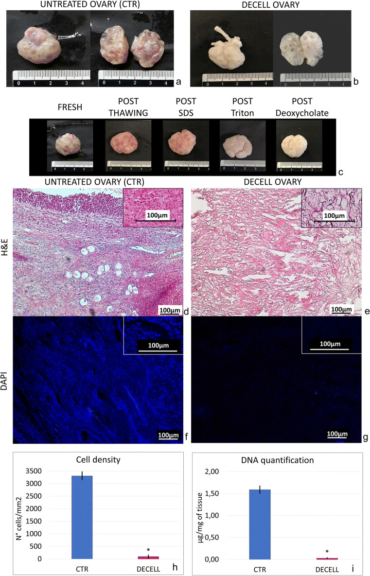
Purpose: To develop a new protocol for whole-ovary decellularization for the production of a 3D bioscaffold suitable for in vitro/ex vivo studies and for the reconstruction of a bioengineered ovary.
Methods: Porcine ovaries were subjected to the decellularization process (DECELL; n = 20) that involved a freeze-thaw cycle, followed by sequential incubations in 0.5% SDS for 3 h, 1% Triton X-100 for 9 h, and 2% deoxycholate for 12 h. Untreated ovaries were used as a control (CTR; n = 6). Both groups were analyzed to evaluate cell and DNA removal as well as ECM preservation. DECELL bioscaffolds were assessed for cytotoxicity and cell homing ability.
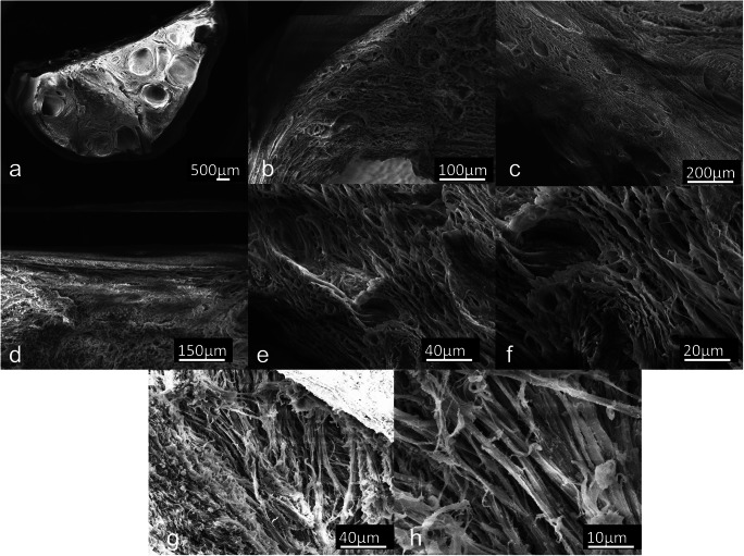
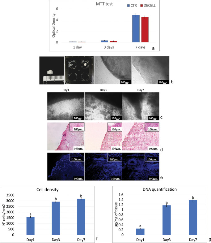
Results: DECELL ovaries maintained shape and homogeneity without any deformation, while their color turned from red to white. Histological staining and DNA quantification confirmed a decrease of 98.11% in DNA content, compared with the native tissue (CTR). Histochemical assessments demonstrated the preservation of intact ECM microarchitecture after the decellularization process. This was also confirmed by quantitative analysis of collagen, elastin, and GAG contents. DECELL bioscaffold showed no cytotoxic effects in co-culture and, when re-seeded with homologous fibroblasts, encouraged a rapid cell adhesion and migration, with repopulating cells increasing in number and aggregating in cluster-like structures, consistent with its ability to sustain cell adherence, proliferation, and differentiation.
Conclusion: The protocol described allows for the generation of a 3D bioscaffold that may constitute a suitable model for ex vivo culture of ovarian cells and follicles, as well as a promising tool for the reconstruction of a bioengineered ovary.
Keywords: 3D bioscaffold; Decellularization; Extracellular matrix; Porcine; Whole ovary.
Conflict of interest statement
The authors declare that they have no conflict of interest.
Figures

Similar articles
-
Creation of a Bioengineered Ovary: Isolation of Female Germline Stem Cells for the Repopulation of a Decellularized Ovarian Bioscaffold.Methods Mol Biol. 2021;2273:139-149. doi: 10.1007/978-1-0716-1246-0_9. Methods Mol Biol. 2021. PMID: 33604850
-
Decellularization of the mouse ovary: comparison of different scaffold generation protocols for future ovarian bioengineering.J Ovarian Res. 2019 Jun 22;12(1):58. doi: 10.1186/s13048-019-0531-3. J Ovarian Res. 2019. PMID: 31228949 Free PMC article.
-
Ovarian Decellularized Bioscaffolds Provide an Optimal Microenvironment for Cell Growth and Differentiation In Vitro.Cells. 2021 Aug 18;10(8):2126. doi: 10.3390/cells10082126. Cells. 2021. PMID: 34440895 Free PMC article.
-
Current Trends on Bioengineering Approaches for Ovarian Microenvironment Reconstruction.Tissue Eng Part B Rev. 2023 Jun;29(3):260-298. doi: 10.1089/ten.TEB.2022.0171. Epub 2023 Mar 9. Tissue Eng Part B Rev. 2023. PMID: 36355603 Review.
-
The Most Ideal Pancreas Extracellular Matrix as a Platform for Pancreas Bioengineering: Decellularization/Recellularization Protocols.Adv Exp Med Biol. 2021;1345:61-70. doi: 10.1007/978-3-030-82735-9_6. Adv Exp Med Biol. 2021. PMID: 34582014 Review.
Cited by
-
Impact of Aging on the Ovarian Extracellular Matrix and Derived 3D Scaffolds.Nanomaterials (Basel). 2022 Jan 21;12(3):345. doi: 10.3390/nano12030345. Nanomaterials (Basel). 2022. PMID: 35159690 Free PMC article.
-
Perfusion and Ultrasonication Produce a Decellularized Porcine Whole-Ovary Scaffold with a Preserved Microarchitecture.Cells. 2023 Jul 15;12(14):1864. doi: 10.3390/cells12141864. Cells. 2023. PMID: 37508528 Free PMC article.
-
Generation of bovine decellularized testicular bio-scaffolds as a 3D platform for testis bioengineering.Front Bioeng Biotechnol. 2025 Jan 14;12:1532107. doi: 10.3389/fbioe.2024.1532107. eCollection 2024. Front Bioeng Biotechnol. 2025. PMID: 39877269 Free PMC article.
-
Characteristics of a decellularized human ovarian tissue created by combined protocols and its interaction with human endometrial mesenchymal cells.Prog Biomater. 2021 Sep;10(3):195-206. doi: 10.1007/s40204-021-00163-6. Epub 2021 Sep 5. Prog Biomater. 2021. PMID: 34482521 Free PMC article.
-
Enhancement of Vascularization and Ovarian Follicle Survival Using Stem Cells in Cryopreserved Ovarian Tissue Transplantation-A Systematic Review.Biology (Basel). 2024 May 14;13(5):342. doi: 10.3390/biology13050342. Biology (Basel). 2024. PMID: 38785824 Free PMC article. Review.
References
-
- Trinh X-B, Peeters F, Tjalma WAA. The thoughts of breast cancer survivors regarding the need for starting hormone replacement therapy. Eur J Obstet Gynecol Reprod Biol. 2006;124:250–253. - PubMed
-
- Yalcinkaya TM, Sittadjody S, Opara EC. Scientific principles of regenerative medicine and their application in the female reproductive system. Maturitas. 2014;77:12–19. - PubMed
MeSH terms
Substances
Grants and funding
LinkOut - more resources
Full Text Sources
Other Literature Sources
