. 2020 Jul;29(7):1629-1640.
doi: 10.1002/pro.3877. Epub 2020 May 25.

Oligomerization of a symmetric β-trefoil protein in response to folding nucleus perturbation

Affiliations

Oligomerization of a symmetric β-trefoil protein in response to folding nucleus perturbation

Connie A Tenorio et al. Protein Sci. 2020 Jul.

Abstract

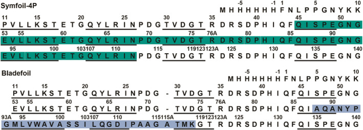
Gene duplication and fusion events in protein evolution are postulated to be responsible for the common protein folds exhibiting internal rotational symmetry. Such evolutionary processes can also potentially yield regions of repetitive primary structure. Repetitive primary structure offers the potential for alternative definitions of critical regions, such as the folding nucleus (FN). In principle, more than one instance of the FN potentially enables an alternative folding pathway in the face of a subsequent deleterious mutation. We describe the targeted mutation of the carboxyl-terminal region of the (internally located) FN of the de novo designed purely-symmetric β-trefoil protein Symfoil-4P. This mutation involves wholesale replacement of a repeating trefoil-fold motif with a "blade" motif from a β-propeller protein, and postulated to trap that region of the Symfoil-4P FN in a nonproductive folding intermediate. The resulting protein (termed "Bladefoil") is shown to be cooperatively folding, but as a trimeric oligomer. The results illustrate how symmetric protein architectures have potentially diverse folding alternatives available to them, including oligomerization, when preferred pathways are perturbed.

Keywords: domain-swapping; folding pathway; protein evolution; protein symmetry.


Conflict of interest statement

M. B. is a cofounder and has equity ownership in Trefoil Therapeutics, Inc.

Figures

FIGURE 1
FIGURE 1
The trefoil‐fold motif within the Symfoil β‐trefoil protein and the site of the “blade” motif substitution. (a) Ribbon diagram of the Symfoil‐4P protein (RCSB accession 3O4D). The green shading identifies the repeating 42‐mer “trefoil‐fold” motif (residue positions 11–52 in Figure 2). (b) Rotation around the vertical threefold axis of symmetry. The green shading identifies a circularly‐permuted definition of the repeating trefoil‐fold motif (residue positions 85–123 in Figure 2). (c) The ab initio predicted structure (green shading) of residues 85–123 highlighted in (b) overlaid onto a “blade” motif (residues 215–248; blue shading) of the β‐propeller protein methylamine dehydrogenase (RCSB accession 1MDA). See Figure 2 for primary sequence alignment
FIGURE 2
FIGURE 2
Primary structure of Symfoil‐4P and BF proteins. Upper panel: the primary structure of Symfoil‐4P with β‐strand secondary structure underlined3, 13 and the folding nucleus region shaded in green. 11 Lower panel: the primary structure of the designed BF protein. The blue shaded region indicates the internal substitution of residue positions 217–246 of the β‐propeller protein methylamine dehydrogenase (RCSB accession 1MDA). This region corresponds to an integral “blade” motif of this seven‐stranded β‐propeller architecture. Underlined regions indicate the β‐strand secondary structure from the 1MDA structure. This substitution should disrupt the C‐terminus region of the Symfoil‐4P folding nucleus (i.e., four of the seven β‐strands in the FN). Alternative forms of the folding nucleus may be possible due to the primary structure symmetry
FIGURE 3
FIGURE 3
Analytical SEC of BF proteins. Upper panel: The BF protein was resolved with analytical SEC on Superdex 200 under both neutral and acidic pH conditions. The effects of salting‐in ((NH4)2SO4) and salting‐out (i.e., denaturing) (GuHCl) Hoffmeister salts at neutral pH were also evaluated. SEC analysis of the Symfoil‐4P control protein under neutral and acidic conditions is also included. The resolution of protein standards used for mass calibration (light gray) are also shown (see Section 4 for protein composition). Lower panel: BF3x and BF4x proteins were analyzed by analytical SEC under acidic (pH 3.0) conditions. Also included are the BF and Symfoil‐4P control proteins. Mass standards (light gray) are also indicated. BF, Bladefoil; SEC, size exclusion chromatography
FIGURE 4
FIGURE 4
Analysis of AUC data of Symfoil‐4P and BF proteins at pH 3.0. The G(s) distributions of BF and Symfoil‐4P proteins from the van Holde–Weischet analysis are shown. Both Symfoil‐4P and BF proteins appear homogeneous, and BF exhibits a sedimentation coefficient that is a 3x stoichiometric ratio of Symfoil‐4P. AUC, analytical ultracentrifugation; BF, Bladefoil
FIGURE 5
FIGURE 5
Isothermal equilibrium denaturation of BF and Symfoil proteins. (a) The effect of pH 11.0–3.0 upon IED of BF protein by GuHCl and monitored by fluorescence (all samples analyzed at 4.37 μM, or 0.070 mg/ml). (b) Effect of gene duplication (3x or 4x) of the BF sequence upon the IED data at pH 3.0. The BF, BF3x and BF4x proteins were analyzed at 4.37 μM, 1.46 μM and 1.09 μM, respectively (i.e., maintaining a constant mass concentration of 0.070 mg/ml). (c) IED of Symfoil‐4P, Difoil and Monofoil proteins at 0.070 mg/ml and analyzed at pH 7.0 and pH 3.0. IED, isothermal equilibrium denaturation
FIGURE 6
FIGURE 6
IED ΔG U versus [GuHCl] for BF proteins at pH 3.0. The BF proteins and IED models utilized in data analysis are indicated. Also shown are the [GuHCl] at the respective ΔG = 0 values. The Symfoil‐4P protein is also included for reference. BF, Bladefoil; IED, isothermal equilibrium denaturation
FIGURE 7
FIGURE 7
DSC endotherm of BF in 0.1 M citrate pH 3.0. Heat capacity is indicated by tick marks in increments of 10 kJ mol−1 K−1. The experimental data (black) is overlaid with a fit to a trimer dissociation model (see Section 4). Fitted thermodynamic values are given in Table 2. DSC, differential scanning calorimetry
FIGURE 8
FIGURE 8
Monomer and oligomeric assemblies for BF proteins. (a) The trimeric assembly of the 42‐mer Symfoil‐4P trefoil‐fold motif (“Monofoil”).3, 13 Solid lines indicate intermolecular interfaces between oligomeric subunits. (b) Representation of the Symfoil‐4P protein (a triplet repeat of the Monofoil 42‐mer sequence). Symfoil‐4P folds as a monomer, forming an overall β‐trefoil architecture comprised of three repeating trefoil‐fold structural motifs. The dashed lines represent intramolecular interfaces between repeating motifs. (c) Trimer assembly of the BF protein consistent with SEC and IED data. The blade motif insertion (gray) disrupts a major portion of the interior FN region; however, sequence symmetry provides a potential alternative FN definition similar to the integral Monofoil sequence (and able to generate in integral b‐trefoil architecture via trimerization). (d) Representation of the BF3x construct and its monomeric assembly that shares properties with the BF trimer assembly. (e) Representation of the BF4x construct. The initial three repeats fold as a monomer like BF3x, and the last repeat provides for trimeric assembly as with BF (consistent with SEC and IED data). The green and blue shading represent instances of the BF4x protein in the hypothesized trimeric assembly. BF, Bladefoil; FN, folding nucleus; IED, isothermal equilibrium denaturation; SEC, size exclusion chromatography

Similar articles

Cited by

References

    1. McLachlan AD. Three‐fold structural pattern in the soybean trypsin inhibitor (Kunitz). J Mol Biol. 1979;133:557–563. - PubMed
    1. Murzin AG, Lesk AM, Chothia C. β‐Trefoil fold. Patterns of structure and sequence in the kunitz inhibitors interleukins‐1β and 1α and fibroblast growth factors. J Mol Biol. 1992;223:531–543. - PubMed
    1. Lee J, Blaber SI, Dubey VK, Blaber M. A polypeptide “building block” for the ß‐trefoil fold identified by “top‐down symmetric deconstruction”. J Mol Biol. 2011;407:744–763. - PubMed
    1. Broom A, Doxey AC, Lobsanov YD, et al. Modular evolution and the origins of symmetry: Reconstruction of a three‐fold symmetric globular protein. Structure. 2012;20:1–11. - PubMed
    1. Voet ARD, Noguchi H, Addy C, et al. Computational design of a self‐assembling symmetrical β‐propeller protein. Proc Natl Acad Sci U S A. 2014;111:15102–15107. - PMC - PubMed

Publication types

Substances