. 2020 Jun 22;375(1801):20190408.
doi: 10.1098/rstb.2019.0408. Epub 2020 May 4.

Chloroplast-to-nucleus retrograde signalling controls intercellular trafficking via plasmodesmata formation

Affiliations

Chloroplast-to-nucleus retrograde signalling controls intercellular trafficking via plasmodesmata formation

Elena E Ganusova et al. Philos Trans R Soc Lond B Biol Sci. .

Abstract

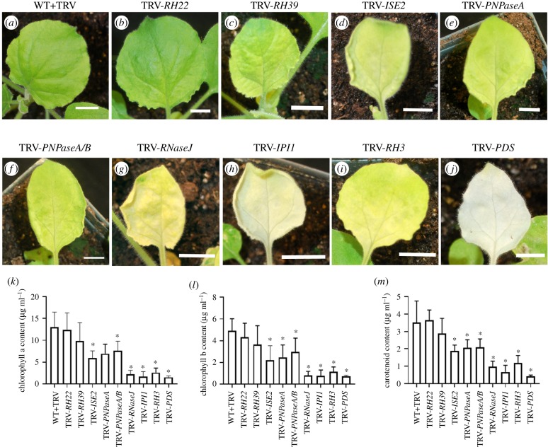
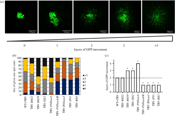
The signalling pathways that regulate intercellular trafficking via plasmodesmata (PD) remain largely unknown. Analyses of mutants with defects in intercellular trafficking led to the hypothesis that chloroplasts are important for controlling PD, probably by retrograde signalling to the nucleus to regulate expression of genes that influence PD formation and function, an idea encapsulated in the organelle-nucleus-PD signalling (ONPS) hypothesis. ONPS is supported by findings that point to chloroplast redox state as also modulating PD. Here, we have attempted to further elucidate details of ONPS. Through reverse genetics, expression of select nucleus-encoded genes with known or predicted roles in chloroplast gene expression was knocked down, and the effects on intercellular trafficking were then assessed. Silencing most genes resulted in chlorosis, and the expression of several photosynthesis and tetrapyrrole biosynthesis associated nuclear genes was repressed in all silenced plants. PD-mediated intercellular trafficking was changed in the silenced plants, consistent with predictions of the ONPS hypothesis. One striking observation, best exemplified by silencing the PNPase homologues, was that the degree of chlorosis of silenced leaves was not correlated with the capacity for intercellular trafficking. Finally, we measured the distribution of PD in silenced leaves and found that intercellular trafficking was positively correlated with the numbers of PD. Together, these results not only provide further support for ONPS but also point to a genetic mechanism for PD formation, clarifying a longstanding question about PD and intercellular trafficking. This article is part of the theme issue 'Retrograde signalling from endosymbiotic organelles'.

Keywords: chloroplast; gun; intercellular trafficking; plasmodesmata; retrograde signalling; tetrapyrrole.


Conflict of interest statement

We declare we have no competing interests.

Figures

Figure 1.
Figure 1.
Phenotypes of silenced plants and their photosynthetic pigment content. (aj) Representative leaves from silenced plants. Silenced gene is shown above figure. WT N. benthamiana infected with TRV virus only (a). (bk) Plants with silenced RH22 (b), RH39 (c), ISE2 (d), PNPaseA (e), PNPaseA/B (f), RNaseJ (g), IPI1 (h), RH3 (i), and PDS (j). Silencing of PDS results in severely chlorotic tissue. Chlorophyll a and b content of silenced plants (k,l), and total carotenoid content of silenced plants (m). Asterisk denotes statistical significance, Student's t-test p < 0.05.
Figure 2.
Figure 2.
Measurement of PD-mediated intercellular trafficking in silenced plants. PD-mediated trafficking was measured by counting the layers of epidermal cells to which GFP spread. At least 10 foci were scored for each of three biological replicates. (a) Representative images depicting GFP trafficking between contiguous layers of epidermal cells. The presence of GFP in cell nuclei was used to determine the presence of GFP in a cell. The cell labelled ‘0' showed expression of GFP in the primary transformed cell but no GFP was seen in surrounding epidermal layers. In the image labelled ‘1', GFP expression was observed in the primary transformed cell and in the nuclei of cells immediately adjacent to the transformed cell only. For subsequent images, the number below the image corresponds to the number of layers away from the primary transformed cell that showed GFP accumulation in the nucleus. Scale bar = 100 µm. (b) Qualitative representation of GFP trafficking data showing the distribution of foci sizes observed for each silenced plant. (c) Layers were assigned values (0 layers = 0, 1 layer = 1, 2 layers = 2, 3 layers = 3, greater than 3 layers = 4) and the median value is shown. Statistical significance was determined by the Mann–Whitney U-test. *p < 0.05.
Figure 3.
Figure 3.
Silencing of genes involved in chloroplast gene expression results in altered expression of CRS target genes. Relative normalized expression of GLK1, LHCB1.2, LHCB2.1, RBCS1A and XTH5 of N. benthamiana in non-silenced TRV-infected controls (WT+TRV) versus (a) TRV-RH22, (b) TRV-RH39, (c) TRV-ISE2, (d) TRV-PNPaseA, (e) TRV-PNPaseA/B, (f) TRV-RNaseJ, (g) TRV-IPI1, and (h) TRV-RH3 silenced plants. Expression was normalized against non-silenced TRV-infected controls and relative expression was calculated using GAPDH and EF1α as reference genes. Data represent mean (± s.e.) from three technical replicates of a representative biological replicate. Statistical analysis was performed by Student's t-test. *p < 0.05. (i) Heatmap showing expression levels of CRS marker genes (GLK1, LHCB1.2, LHCB2.1, RBCS1A and XTH5) in silenced plants. The rows represent CRS marker genes and the columns represent silenced genes. Scale is shown on right.
Figure 4.
Figure 4.
Silencing of genes involved in chloroplast gene expression results in changes in expression of genes of the tetrapyrrole biosynthesis pathway. qPCR analysis of expression of HEMA1, GUN2, 3, 4 and 5 in non-silenced TRV-infected controls (WT+TRV) and (a) TRV-RH22, (b) TRV-RH39, (c) TRV-ISE2, (d) TRV-PNPaseA, (e) TRV-PNPaseA/B, (f) TRV-RNaseJ, (g) TRV-IPI1, and (h) TRV-RH3 silenced plants. GAPDH and EF1α were used as reference genes for calculating the relative expression and normalized with TRV infected wild-type (WT+TRV). The data are presented as the mean (±s.e.) from three technical replicates of a representative biological replicate. Statistical analysis was performed by Student's t-test. *p < 0.05. (i) Heatmap showing expression levels of HEMA1 and the GUN genes in silenced plants. Scale is on right.
Figure 5.
Figure 5.
Quantification of PD density in silenced plants. PD in pitfields were labelled with the general PD marker PDLP1-GFP. Representative maximum projections of fields of view showing PDLP1-GFP-labelled pitfields in (a) WT+TRV, (b) TRV-PNPaseA (increased number of pitfields), and (c) TRV-IPI1 (decreased numbers of pitfields) plants. (d) Individual images (confocal planes 3–14 of a confocal stack) from z-stacks used to generate maximum projections (MP) showing the distribution of pitfields in the z-axis. Images correspond to the region outlined in white in (a). Scale bar = 10 µm. (e) PD density scores were calculated for each image from three biological replicates (rep. 1 (blue squares), rep. 2 (yellow circles) and rep. 3 (red diamonds). See Material and methods for more details. Statistical significance was determined by Mann–Whitney U-test. *p < 0.0001. (f) Linear regression curve showing the positive correlation of PD density score with layers of GFP movement. The average PD density score and the median layers of movement across the three biological replicates were used to generate the graph.
Figure 6.
Figure 6.
Model for the control of formation of secondary PD by CRS. Primary PD (1° PD) are formed during cell division while secondary PD (2° PD) arise in the absence of cell division. Under normal control conditions or in WT plants with normal RNA processing (a), CRS (black arrow) regulates the expression of PDANGs that are involved in the formation of secondary PD. Disruption of chloroplast RNA processing and gene expression, as in the silenced plants used in this study, leads to defects in chloroplast development and function, causing altered CRS (red arrow) which results in changes in the expression of PDANGs (b). These changes result in increased (I) or decreased (II) secondary PD formation.

References

    1. Lucas WJ, Lee JY. 2004. Plasmodesmata as a supracellular control network in plants. Nat. Rev. Mol. Cell Biol. 5, 712–726. (10.1038/nrm1470) - DOI - PubMed
    1. Patrick JW. 2013. Does Don Fisher's high-pressure manifold model account for phloem transport and resource partitioning? Front. Plant Sci. 4, 184 (10.3389/fpls.2013.00184) - DOI - PMC - PubMed
    1. Oparka KJ, et al. 1999. Simple, but not branched, plasmodesmata allow the nonspecific trafficking of proteins in developing tobacco leaves. Cell 97, 743–754. (10.1016/S0092-8674(00)80786-2) - DOI - PubMed
    1. Wu SW, Kumar R, Iswanto ABB, Kim JY. 2018. Callose balancing at plasmodesmata. J. Exp. Bot. 69, 5325–5339. (10.1093/jxb/ery317) - DOI - PubMed
    1. Amsbury S, Kirk P, Benitez-Alfonso Y. 2017. Emerging models on the regulation of intercellular transport by plasmodesmata-associated callose. J. Exp. Bot. 69, 105–115. (10.1093/jxb/erx337) - DOI - PubMed

Publication types

MeSH terms

Substances

LinkOut - more resources