. 2007 Jul;16(3):135-145.
doi: 10.1053/j.jepm.2007.06.002. Epub 2007 Sep 18.

Rabbit Clinical Pathology

Affiliations

Rabbit Clinical Pathology

Alessandro Melillo. J Exot Pet Med. 2007 Jul.

Abstract

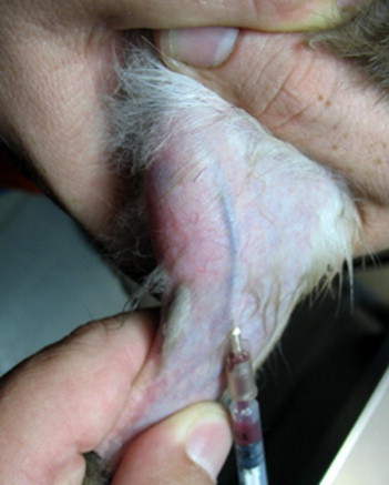
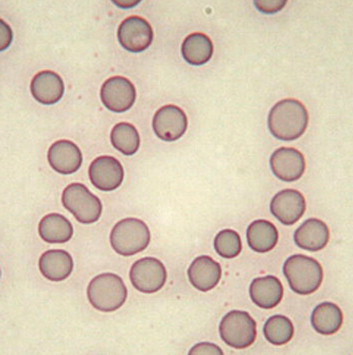
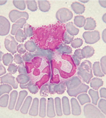
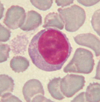
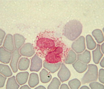
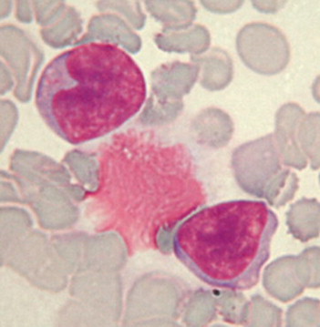
With rabbit patients, as in other species, analyzing blood and urine samples can be useful and informative, although interpretation of the results is sometimes challenging. This article summarizes the interpretation of laboratory results from rabbits. Hematological parameters can yield information about the red blood cell population and leukocyte response to stress and pathogens. Biochemistry evaluation can be used to investigate liver, kidney, and other organ function, and urinalysis results may yield additional information about kidney function and electrolyte imbalances. Serological tests are available for several pathogens of rabbits, including Encephalitozoon cuniculi, although the significance of positive results and antibody titers is not clear. Serum protein electrophoresis aids the understanding of protein disorders and the immune response to acute and chronic inflammation.

Keywords: biochemistry; blood sampling; hematology; rabbit; serum protein electrophoresis; urinalysis.


Figures

Figure 1
Figure 1
Blood collection from the saphenous vein.
Figure 2
Figure 2
Anisocytosis and a Howell-Jolly body. In domestic carnivores, anisocytosis usually reflects the presence of reticulocytes and indicates regenerative anemia. This is not true in rabbits in which 1% to 4% of the circulating erythrocytes may be reticulocytes. The occasional Howell-Jolly body is not clinically significant either.
Figure 3
Figure 3
Neutrophils. Rabbit neutrophils are rounded cells with a diameter of 10 to 15 μm. The nucleus of rabbit neutrophils is distinctly segmented and stained a deep purple-blue; the segments are connected by thin strands of chromatin. In the clear cytoplasm, variable numbers of granules can be seen, ranging in size and color (small and pink or large and reddish). The relative number of each granule population may alter the staining characteristics of the cell. In some cases, laboratories report rabbit neutrophils as heterophils or acidophils because of the deep pink to red stained granules in the cells. Regardless of how the cells are labeled, there is no functional difference between the cells.
Figure 4
Figure 4
Large lymphocyte. Rabbits have 2 distinct populations of lymphocytes: small (7-10 μm) and large (10-15 μm). Lymphocytes are round cells with a round or slightly oval deep purple nucleus. Cytoplasm is scarce or absent in the small lymphocytes and more abundant and bluish in the large ones. Some large lymphocytes may present a clear halo around the nucleus and even a few azurophilic granules.
Figure 5
Figure 5
Eosinophil. Rabbit eosinophils measure 10 to 16 μm in diameter and have a purple bilobed or horseshoe-shaped nucleus. The cytoplasm is obscured by so many granules that the cell looks orange-pink and foamy. The abundance of granules is the main difference between the eosinophils and the neutrophils. Removal of histamine and histamine-like toxins is the most important function of the eosinophils, suggesting that they therefore play an important role in controlling allergic reactions.
Figure 6
Figure 6
Monocytes. Monocytes are the largest of the leukocytes, with an average diameter of 15 to 18 μm. Their purple nucleus is usually lobulated, but may be horseshoe or bean shaped. Monocytes have more cytoplasm than lymphocytes, and their cytoplasm stains a blue-gray mottled color.

References

    1. Harcourt-Brown F.M., Baker S.J. Parathyroid hormone, hematological and biochemical parameters in relation to dental disease and husbandry in pet rabbits. J Small Anim Pract. 2001;42:130–136. - PubMed
    1. Fudge A.M. Rabbit hematology. In: Fudge A.M., editor. Laboratory Medicine: Avian and Exotic Pets. WB Saunders Company; Philadelphia, PA: 2000. pp. 273–275.
    1. Saunders R.A., Davies R.R. Blackwell Publishing; Oxford, UK: 2005. Notes on Rabbit Internal Medicine.
    1. Weisbroth S.H. Neoplastic diseases. In: Manning P.J., Ringler D.H., Newcomer C.E., editors. The Biology of Laboratory Rabbit. (ed 2) Academic Press; New York, NY: 1994. pp. 259–292.
    1. Toth L.A., Krueger J.M. Alteration of sleep in rabbits by Staphylococcus aureus infection. Infect Immun. 1988;56:1785–1791. - PMC - PubMed

LinkOut - more resources