Meta-Analysis
. 2020 Apr 17:11:203.
doi: 10.3389/fendo.2020.00203. eCollection 2020.

Early Endothelial Dysfunction in Type 1 Diabetes Is Accompanied by an Impairment of Vascular Smooth Muscle Function: A Meta-Analysis

Affiliations
Meta-Analysis

Early Endothelial Dysfunction in Type 1 Diabetes Is Accompanied by an Impairment of Vascular Smooth Muscle Function: A Meta-Analysis

Elodie Lespagnol et al. Front Endocrinol (Lausanne). .

Abstract

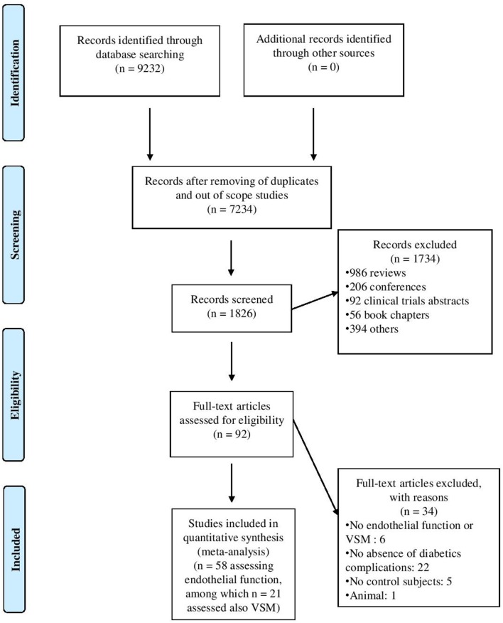
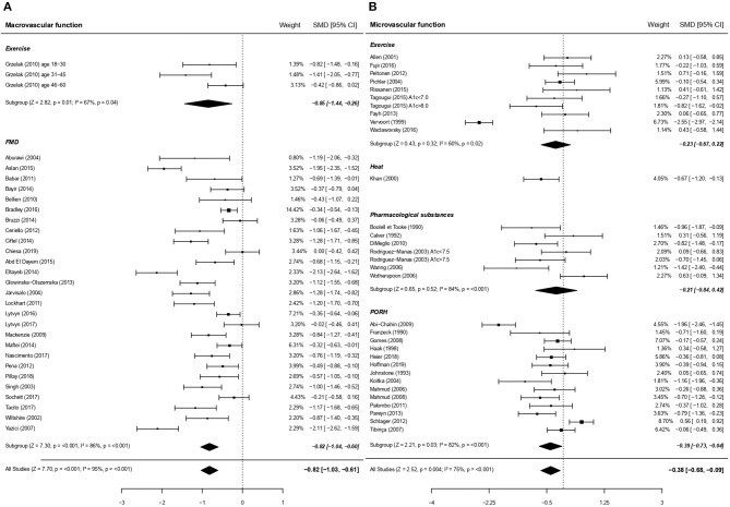
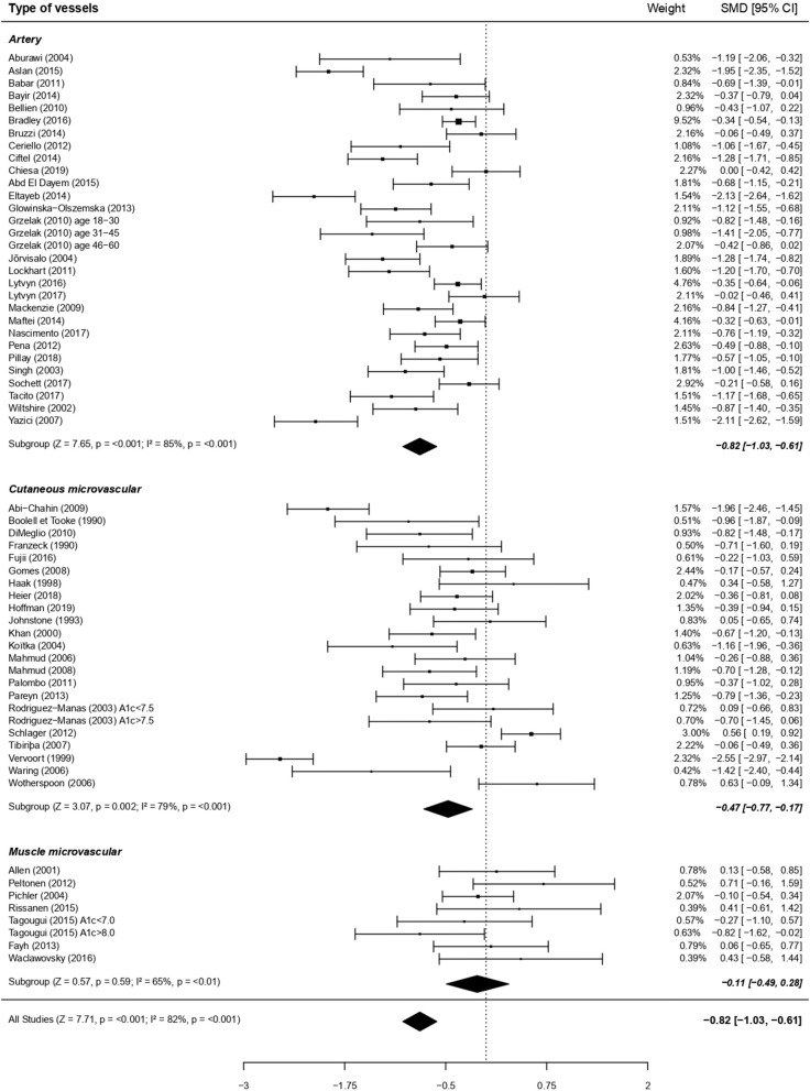
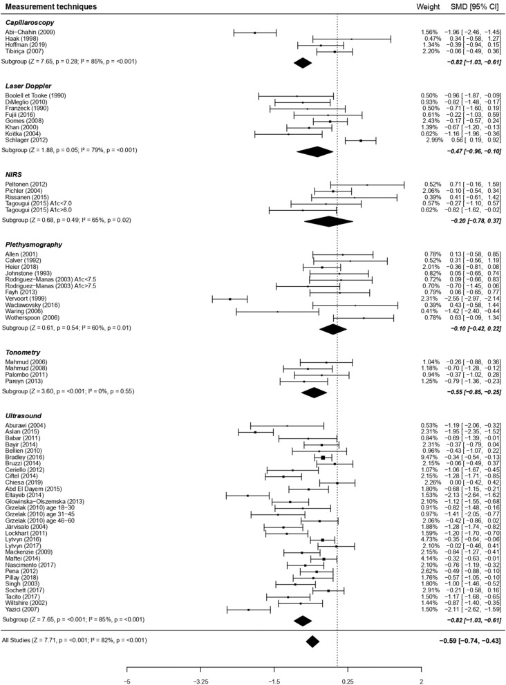
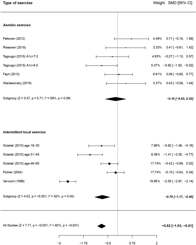
Background: A large yet heterogeneous body of literature exists suggesting that endothelial dysfunction appears early in type 1 diabetes, due to hyperglycemia-induced oxidative stress. The latter may also affect vascular smooth muscles (VSM) function, a layer albeit less frequently considered in that pathology. This meta-analysis aims at evaluating the extent, and the contributing risk factors, of early endothelial dysfunction, and of the possible concomitant VSM dysfunction, in type 1 diabetes. Methods: PubMed, Web of Sciences, Cochrane Library databases were screened from their respective inceptions until October 2019. We included studies comparing vasodilatory capacity depending or not on endothelium (i.e., endothelial function or VSM function, respectively) in patients with uncomplicated type 1 diabetes and healthy controls. Results: Fifty-eight articles studying endothelium-dependent function, among which 21 studies also assessed VSM, were included. Global analyses revealed an impairment of standardized mean difference (SMD) (Cohen's d) of endothelial function: -0.61 (95% CI: -0.79, -0.44) but also of VSM SMD: -0.32 (95% CI: -0.57, -0.07). The type of stimuli used (i.e., exercise, occlusion-reperfusion, pharmacological substances, heat) did not influence the impairment of the vasodilatory capacity. Endothelial dysfunction appeared more pronounced within macrovascular than microvascular beds. The latter was particularly altered in cases of poor glycemic control [HbA1c > 67 mmol/mol (8.3%)]. Conclusions: This meta-analysis not only corroborates the presence of an early impairment of endothelial function, even in response to physiological stimuli like exercise, but also highlights a VSM dysfunction in children and adults with type 1 diabetes. Endothelial dysfunction seems to be more pronounced in large than small vessels, fostering the debate on their relative temporal appearance.

Keywords: endothelial function; exercise; macrocirculation; microcirculation; peripheral vascular disease; smooth muscle function; type 1 diabetes.


Figures

Figure 1
Figure 1
Flow-chart outlining the process of study selection.
Figure 2
Figure 2
Forest plots of studies on endothelial function, according to size of vessels studied, in subjects with type 1 diabetes vs. controls. (A) Forest plot of studies on endothelial function focusing on macrovascular function. (B) Forest plot of studies on endothelial function focusing on microvascular function. In these figures the priority was set on macrocirculation over microcirculation. Similar results are obtained when priority is placed on microcirculation (SMD −0.59; 95% CI −0.74, −0.43, P < 0.001). PORH, Post-occlusive reactive hyperaemia; FMD, Flow Mediated Dilation. For each subgroup, the following values are indicated: Z- and p-value; I2, heterogeneity and p for heterogeneity. When comparing macro and microcirculation with metaregressions (for qualitative data), the difference was significant (moderator P = 0.001) with a greater dysfunction due to type 1 diabetes for macrovessels.
Figure 3
Figure 3
Forest plots of studies on endothelial function, according to types of vessels studied, in subjects with type 1 diabetes vs. controls. For each subgroup, the following values are indicated: Z and p-value; I2, heterogeneity and p for heterogeneity. When comparing the three vessels types with metaregressions (for qualitative data), the difference was significant (moderator P = 0.005), with a greater dysfunction for artery compared to muscle microvessels (post-hoc comparison using Bonferroni correction, p < 0.05) and a tendency for a greater dysfunction at artery compared to cutaneous microvessels (post-hoc comparison using Bonferroni correction, P = 0.09).
Figure 4
Figure 4
Forest plots of studies on endothelial function, according to measurement techniques, in subjects with type 1 diabetes vs. controls. For each subgroup, the following values are indicated: Z and p-value; I2, heterogeneity and p for heterogeneity. When comparing the six types of techniques with metaregressions (for qualitative data), the difference was significant (moderator P = 0.02), with a more visible dysfunction when using ultrasounds vs. plethysmography (post-hoc comparison using Bonferroni correction, P < 0.01).
Figure 5
Figure 5
Forest plots of studies on endothelial function in response to exercise, according to the type of exercise, in subjects with type 1 diabetes vs. controls. For each subgroup, the following values are indicated: Z and p-value; I2, heterogeneity and p for heterogeneity. When comparing the two types of exercise with metaregressions (for qualitative data), the difference was significant (moderator P = 0.04), with a more visible dysfunction when using intermittent local exercise vs. aerobic exercise.
Figure 6
Figure 6
Forest plot of studies on VSM function in subjects with type 1 diabetes vs. controls. For each subgroup, the following values are indicated: Z and p-value; I2, heterogeneity and p for heterogeneity. There was no difference between macro and microcirculation (with metaregression for qualitative data: moderator P = 0.60).

Similar articles

Cited by

References

    1. Nathan DM, Cleary PA, Backlund JY, Genuth SM, Lachin JM, Orchard TJ, et al. . Intensive diabetes treatment and cardiovascular disease in patients with type 1 diabetes. N Engl J Med. (2005) 353:2643–53. 10.1056/NEJMoa052187 - DOI - PMC - PubMed
    1. Jarvisalo MJ, Raitakari M, Toikka JO, Putto-Laurila A, Rontu R, Laine S, et al. . Endothelial dysfunction and increased arterial intima-media thickness in children with type 1 diabetes. Circulation. (2004) 109:1750–5. 10.1161/01.CIR.0000124725.46165.2C - DOI - PubMed
    1. Mahmud FH, Earing MG, Lee RA, Lteif AN, Driscoll DJ, Lerman A. Altered endothelial function in asymptomatic male adolescents with type 1 diabetes. Congenit Heart Dis. (2006) 1:98–103. 10.1111/j.1747-0803.2006.00015.x - DOI - PubMed
    1. Wink DA, Miranda KM, Espey MG, Pluta RM, Hewett SJ, Colton C, et al. . Mechanisms of the antioxidant effects of nitric oxide. Antioxid Redox Signal. (2001) 3:203–13. 10.1089/152308601300185179 - DOI - PubMed
    1. Quyyumi AA. Endothelial function in health and disease: new insights into the genesis of cardiovascular disease. Am J Med. (1998). 105:32S−9S. 10.1016/S0002-9343(98)00209-5 - DOI - PubMed