

Comparison of the diagnostic methods on the canine adenovirus type 2 infection
- PMID: 32362936
- PMCID: PMC7188356
- DOI: 10.1111/j.1755-9294.2010.01073.x
Comparison of the diagnostic methods on the canine adenovirus type 2 infection
Abstract
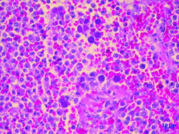
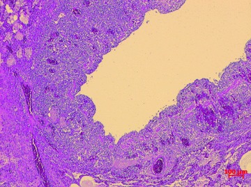
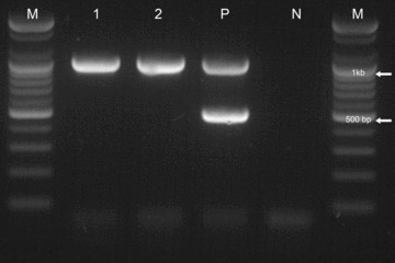
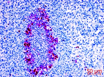
Background and aims: Canine adenovirus type 2 (CAV-2) infection is typically diagnosed histopathologically since intranuclear inclusion bodies (IN/IBs) are demonstrable in the infected lung. However, it is sometimes difficult to identify IN/IBs, particularly in autolyzed tissues or samples from both early and late stages of infection, and other methods were presently developed. Methods: Stray dog samples were evaluated by histopathology, polymerase chain reaction (PCR), and immunohistochemistry (IHC) to investigate the status of the CAV-2 infection on the stray dogs in Korea. Histologic tests were performed, and dogs with pneumonic lungs were further evaluated by IHC and PCR. Results: Pathognomonic IN/IBs were identified in 3 of 213 lungs; CAV-2 PCR was positive for 27 of 213 pneumonic lungs. A total of 7 of 27 CAV-2 PCR-positive lungs were IHC-positive. No PCR-negative lung was IHC-positive. Positive results were primarily detected in the IN/IBs of the bronchial epithelial cells, macrophages, and very rarely in the cytoplasm of bronchial epithelial cells. Conclusions: IHC was a more reliable diagnostic method than conventional pathologic methods in the present study, and suggests that IHC should be routinely used in the diagnosis of CAV-2 infection. Further, PCR alone may not be adequate for CAV-2 diagnosis.
Keywords: CAV‐2; PCR; canine adenovirus type 2; immunohistochemistry; pathology.
© 2010 The Korean Society for Cytopathology, The Korean Society for Legal Medicine, The Korean Society of Oral and Maxillofacial Pathology, The Korean Society of Pathologists, The Korean Society of Toxicological Pathology, The Korean Society of Veterinary Pathology and Blackwell Publishing Asia Pty Ltd.
Figures
Similar articles
-
Use of polymerase chain reaction and immunohistochemistry for detection of canine adenovirus type 1 in formalin-fixed, paraffin-embedded liver of dogs with chronic hepatitis or cirrhosis.J Vet Diagn Invest. 1998 Oct;10(4):320-5. doi: 10.1177/104063879801000402. J Vet Diagn Invest. 1998. PMID: 9786518
-
High prevalence of antibodies against canine adenovirus (CAV) type 2 in domestic dog populations in South Africa precludes the use of CAV-based recombinant rabies vaccines.Vaccine. 2013 Aug 28;31(38):4177-82. doi: 10.1016/j.vaccine.2013.06.089. Epub 2013 Jul 16. Vaccine. 2013. PMID: 23867013
-
Simultaneous canine distemper virus, canine adenovirus type 2, and Mycoplasma cynos infection in a dog with pneumonia.Vet Pathol. 2007 Jul;44(4):508-12. doi: 10.1354/vp.44-4-508. Vet Pathol. 2007. PMID: 17606512
-
[Problems and limitations of conventional and innovative methods for the diagnosis of Toxoplasmosis in humans and animals].Parassitologia. 2004 Jun;46(1-2):177-81. Parassitologia. 2004. PMID: 15305712 Review. Italian.
-
Canine Adenovirus 2: A Natural Choice for Brain Circuit Dissection.Front Mol Neurosci. 2020 Feb 27;13:9. doi: 10.3389/fnmol.2020.00009. eCollection 2020. Front Mol Neurosci. 2020. PMID: 32174812 Free PMC article. Review.
Cited by
-
Development of indirect ELISA for the detection of canine adenovirus type 2 antibodies in dog sera.J Vet Sci. 2020 Jul;21(4):e63. doi: 10.4142/jvs.2020.21.e63. J Vet Sci. 2020. PMID: 32735100 Free PMC article.
-
Siewert-Kartagener's syndrome in a dog.J Vet Sci. 2023 Jul;24(4):e57. doi: 10.4142/jvs.23029. J Vet Sci. 2023. PMID: 37532300 Free PMC article.
-
Canine Adenoviruses in Wildlife: Role in At-Risk Species Conservation and Interface with Domestic Animals.Pathogens. 2025 Feb 18;14(2):200. doi: 10.3390/pathogens14020200. Pathogens. 2025. PMID: 40005575 Free PMC article. Review.
-
Immunogenicity of a new, inactivated canine adenovirus type 2 vaccine for dogs.Clin Exp Vaccine Res. 2020 Jan;9(1):40-47. doi: 10.7774/cevr.2020.9.1.40. Epub 2020 Jan 31. Clin Exp Vaccine Res. 2020. PMID: 32095439 Free PMC article.
-
Development of one-step multiplex real-time PCR for the detection of CHV-1, CAdV-2, and CDV.Front Vet Sci. 2025 May 9;12:1583769. doi: 10.3389/fvets.2025.1583769. eCollection 2025. Front Vet Sci. 2025. PMID: 40417353 Free PMC article.
References
-
- Buonavoglia C, Martella V. Canine respiratory viruses. Vet Res 2007; 38: 355–73. - PubMed
-
- Carlton WW, McGavin MD. Thomson's Special Veterinary Pathology. St. Louis: Mosby , 1995; 125–95.
-
- Damián M, Morales E, Salas G, Trigo FJ. Immunohistochemical detection of antigens of distemper, adenovirus and parainfluenza viruses in domestic dogs with pneumonia. J Comp Pathol 2005; 133: 289–93. - PubMed
LinkOut - more resources
Full Text Sources