

First Reported Case of Extramedullary Plasmacytoma of the Appendix
- PMID: 32362968
- PMCID: PMC7188365
- DOI: 10.14740/gr1277
First Reported Case of Extramedullary Plasmacytoma of the Appendix
Abstract
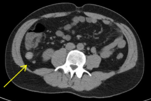
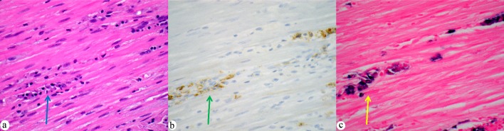
An extramedullary plasmacytoma involving the gastrointestinal tract is extremely rare. We report an appendiceal extramedullary plasmacytoma in a 35-year-old man who presented to the emergency department because of upper abdominal pain. Computed tomography (CT) imaging revealed an incidental mass (3.7 × 1.9 × 1.6 cm) at the tip of the appendix. Microscopically, the appendix, periappendiceal soft tissue, and nearby lymph nodes were diffusely infiltrated by plasma cells that were kappa light chain restricted. Subsequent workup included an unremarkable bone marrow biopsy, as well as urine and serum electrophoresis. A diagnosis of kappa-restricted solitary extramedullary plasmacytoma was made. To our knowledge, this is the first case reported of an appendiceal extramedullary plasmacytoma in the medical literature.
Keywords: Appendix; Extramedullary plasmacytoma; Gastrointestinal tract; Plasma cell neoplasm.
Copyright 2020, Evans et al.
Conflict of interest statement
None to declare.
Figures
Similar articles
-
Solitary Extramedullary Plasmacytoma With Development of T-cell Anaplastic Large-Cell Lymphoma: A Rare Case Report and Literature Review.Cureus. 2023 Apr 26;15(4):e38153. doi: 10.7759/cureus.38153. eCollection 2023 Apr. Cureus. 2023. PMID: 37252473 Free PMC article.
-
Solitary extramedullary plasmacytoma presenting as an adrenal tumor: case report and literature review.Gland Surg. 2021 Mar;10(3):1158-1164. doi: 10.21037/gs-20-773. Gland Surg. 2021. PMID: 33842260 Free PMC article.
-
Unusual presentation of a soft palate mass: A rare case report of solitary extramedullary plasmacytoma.Int J Surg Case Rep. 2021 Feb;79:193-197. doi: 10.1016/j.ijscr.2021.01.033. Epub 2021 Jan 15. Int J Surg Case Rep. 2021. PMID: 33482447 Free PMC article.
-
Solitary extramedullary plasmacytoma of the adrenal gland: a rare case report with review of the literature.Int J Clin Exp Pathol. 2014 Dec 1;7(12):9072-5. eCollection 2014. Int J Clin Exp Pathol. 2014. PMID: 25674290 Free PMC article. Review.
-
Solitary extramedullary plasmacytoma in the lung misdiagnosed as lung cancer: A case report and literature review.Front Oncol. 2022 Aug 30;12:950383. doi: 10.3389/fonc.2022.950383. eCollection 2022. Front Oncol. 2022. PMID: 36110956 Free PMC article. Review.
Cited by
-
Intestinal perforation with abdominal abscess caused by extramedullary plasmacytoma of small intestine: A case report and literature review.World J Gastrointest Surg. 2022 Jun 27;14(6):611-620. doi: 10.4240/wjgs.v14.i6.611. World J Gastrointest Surg. 2022. PMID: 35979418 Free PMC article.
References
-
- Mjoli M, Vorajee N, Naidoo Y, Madiba T. Solitary extramedullary plasmacytoma of the colon, rectum and anus. S Afr J Surg. 2016;54(2):45–47. - PubMed