

Spontaneous Remission in a Patient With Acute Myeloid Leukemia Leading to Undetectable Minimal Residual Disease
- PMID: 32362981
- PMCID: PMC7188378
- DOI: 10.14740/jh606
Spontaneous Remission in a Patient With Acute Myeloid Leukemia Leading to Undetectable Minimal Residual Disease
Abstract
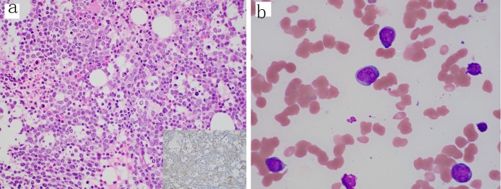
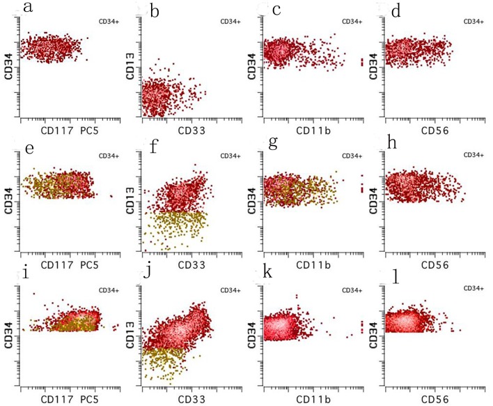
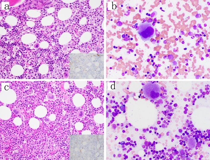
Although rare, spontaneous remission (SR) of acute myeloid leukemia (AML) has been reported in the literature, the underlying mechanisms driving remission remain unknown. However, it is most commonly associated with a preceding severe infection. We present a case of a 40-year-old man with no past medical history who presented to our hospital with severe left hip pain and fevers and was found to have AML. Chemotherapy was delayed because the patient required extensive debridement and fasciotomy of his left hip and a prolonged course of antibiotics. After his acute illness had stabilized, a repeat bone marrow biopsy was performed which showed no abnormal myeloid blasts and resolution of his original cytogenetic and molecular abnormalities. At the time of this writing, our patient remains in remission with undetectable minimal residual disease (MRD), now 14 months from his initial diagnosis of AML.
Keywords: AML; Minimal residual disease; Spontaneous remission.
Copyright 2020, Helbig et al.
Conflict of interest statement
None to declare.
Figures
Similar articles
-
Therapy-related myeloid neoplasm in an 18-year-old boy with B-lymphoblastic leukemia.Exp Mol Pathol. 2017 Dec;103(3):263-266. doi: 10.1016/j.yexmp.2017.11.007. Epub 2017 Nov 16. Exp Mol Pathol. 2017. PMID: 29155023
-
Allogeneic Hematopoietic Cell Transplantation for Acute Myeloid Leukemia: Time to Move Toward a Minimal Residual Disease-Based Definition of Complete Remission?J Clin Oncol. 2016 Feb 1;34(4):329-36. doi: 10.1200/JCO.2015.63.3826. Epub 2015 Dec 14. J Clin Oncol. 2016. PMID: 26668349 Free PMC article.
-
Spontaneous remission in adult acute myeloid leukemia in association with systemic bacterial infection-case report and review of the literature.Ann Hematol. 2004 Mar;83(3):189-94. doi: 10.1007/s00277-003-0741-y. Epub 2003 Oct 3. Ann Hematol. 2004. PMID: 15064869 Review.
-
Increase in myeloid-derived suppressor cells (MDSCs) associated with minimal residual disease (MRD) detection in adult acute myeloid leukemia.Int J Hematol. 2015 Nov;102(5):579-86. doi: 10.1007/s12185-015-1865-2. Epub 2015 Sep 10. Int J Hematol. 2015. PMID: 26358057
-
Detection of minimal residual disease in acute myelogenous leukemia.Acta Haematol. 2004;112(1-2):40-54. doi: 10.1159/000077559. Acta Haematol. 2004. PMID: 15179004 Review.
Cited by
-
Antibiotic and glucocorticoid-induced recapitulated hematological remission in acute myeloid leukemia: A case report and review of literature.World J Clin Cases. 2022 Aug 6;10(22):7890-7898. doi: 10.12998/wjcc.v10.i22.7890. World J Clin Cases. 2022. PMID: 36158489 Free PMC article.
-
Genetic Profiling of Acute and Chronic Leukemia via Next-Generation Sequencing: Current Insights and Future Perspectives.Hematol Rep. 2025 Mar 28;17(2):18. doi: 10.3390/hematolrep17020018. Hematol Rep. 2025. PMID: 40277842 Free PMC article. Review.
-
Always stressed but never exhausted: how stem cells in myeloid neoplasms avoid extinction in inflammatory conditions.Blood. 2023 Jun 8;141(23):2797-2812. doi: 10.1182/blood.2022017152. Blood. 2023. PMID: 36947811 Free PMC article.
-
[A case of spontaneous remission of acute myeloid leukemia with MLL-AF9 rearrangement and abnormal liver function].Zhonghua Xue Ye Xue Za Zhi. 2021 Oct 14;42(10):851-857. doi: 10.3760/cma.j.issn.0253-2727.2021.10.010. Zhonghua Xue Ye Xue Za Zhi. 2021. PMID: 34788926 Free PMC article. Chinese.
-
Transient Spontaneous Remission of Acute Myeloid Leukemia with Mutated RUNX1: A Rare Report.Indian J Hematol Blood Transfus. 2024 Jul;40(3):545-546. doi: 10.1007/s12288-024-01758-2. Epub 2024 Mar 22. Indian J Hematol Blood Transfus. 2024. PMID: 39011264 Free PMC article. No abstract available.
References
-
- Eisenlohr C. Leucaemia lienalis, lymphatica et medullaris mit multiplen Gehirnnervenlahmungen. Archiv fur Pathologie, Anatomie und Physiologie und fur Klinische Medizin. 1878;73:56–73. doi: 10.1007/BF01994747. - DOI
Publication types
Grants and funding
LinkOut - more resources
Full Text Sources
Research Materials
Miscellaneous