Review
. 2020 Apr 17:7:39.
doi: 10.3389/fnut.2020.00039. eCollection 2020.

Feeding Rhythms and the Circadian Regulation of Metabolism

Affiliations
Review

Feeding Rhythms and the Circadian Regulation of Metabolism

Lauren Pickel et al. Front Nutr. .

Abstract

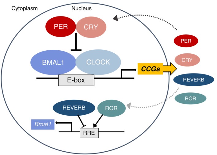
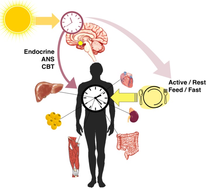
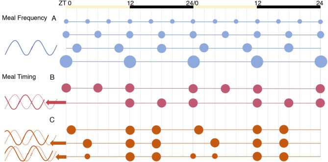
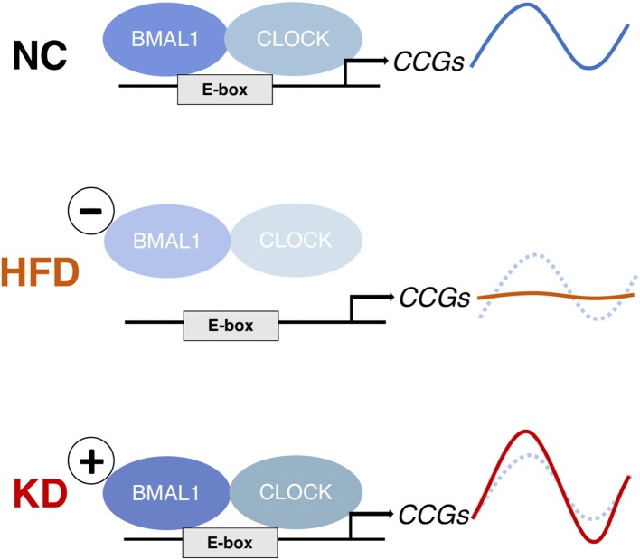
The molecular circadian clock regulates metabolic processes within the cell, and the alignment of these clocks between tissues is essential for the maintenance of metabolic homeostasis. The possibility of misalignment arises from the differential responsiveness of tissues to the environmental cues that synchronize the clock (zeitgebers). Although light is the dominant environmental cue for the master clock of the suprachiasmatic nucleus, many other tissues are sensitive to feeding and fasting. When rhythms of feeding behavior are altered, for example by shift work or the constant availability of highly palatable foods, strong feedback is sent to the peripheral molecular clocks. Varying degrees of phase shift can cause the systemic misalignment of metabolic processes. Moreover, when there is a misalignment between the endogenous rhythms in physiology and environmental inputs, such as feeding during the inactive phase, the body's ability to maintain homeostasis is impaired. The loss of phase coordination between the organism and environment, as well as internal misalignment between tissues, can produce cardiometabolic disease as a consequence. The aim of this review is to synthesize the work on the mechanisms and metabolic effects of circadian misalignment. The timing of food intake is highlighted as a powerful environmental cue with the potential to destroy or restore the synchrony of circadian rhythms in metabolism.

Keywords: circadian; fasting; high fat diet; ketogenic diet; metabolism; peripheral clock; time-restricted feeding.


Figures

Figure 1
Figure 1
The mammalian molecular clock. The complex of BMAL1 and CLOCK rhythmically binds E-box elements to activate the transcription of clock-controlled genes (CCG), including other parts of the core clock which form the negative arm of the feedback loop. The PER and CRY proteins translated from Period (–3) and Cryptochrome (1, 2) translocate to the nucleus and inhibit the activity of CLOCK:BMAL1. Eventually they are tagged for proteasomal degradation and the cycle begins again approximately every 24 h. In a secondary loop, the protein products of the Rev-erbs (α and β) and RORs (α, β, and γ) inhibit and activate Bmal1 transcription, respectively. Other CCGs are involved in the pathways of metabolic regulation. A subset of these (including PPARs, PGC1α, AMPK, and NAD+ dependent SIRT1) in turn also regulate the clock. Figure adapted with permission from Gaucher et al. (12).
Figure 2
Figure 2
Entrainment and alignment of the SCN and peripheral clocks. The primary zeitgeber of the SCN oscillator is light, whereas peripheral clocks are strongly entrained by feeding/fasting rhythms. The SCN synchronizes peripheral rhythms directly through endocrine and autonomic nervous system (ANS) signals and the regulation of the core body temperature (CBT), as well as indirectly through behavioral feedback from rhythms in activity and feeding.
Figure 3
Figure 3
The effects of meal frequency vs. timing on peripheral tissue clocks [from results of (105)]. The phase of Per2 expression was similar in all conditions for the tissues tested: liver, kidney, and submandibular gland. (A) Meal frequency, spreading calories equally into 2, 3, 4, or 6 meals per day, did not alter clock phase. (B) Daytime (inactive phase) feeding significantly advanced phase. (C) Using a three-meal schedule designed to mimic human eating habits, a “late dinner” meal (in the early inactive phase) caused a dramatic phase advance, which was partially reversed by splitting the dinner meal to include a “pre-dinner snack”.
Figure 4
Figure 4
The effects of a high-fat diet (HFD) and ketogenic diet (KD) on clock-controlled gene (CCG) expression in mouse liver. The HFD abolishes rhythms in CCGs by reducing CLOCK:BMAL1 chromatin binding (136), whereas the KD increases rhythmic CLOCK:BMAL1 binding and thereby CCG expression amplitude (147). Both diets induce de novo rhythms in other metabolic genes because of the rhythmic nuclear accumulation of non-clock transcription regulators (PPARg and SREBP-1 in liver under HFD; PPARα in intestine and rhythmic histone deacetylation by serum BHB under KD).

References

    1. Patel VR, Ceglia N, Zeller M, Eckel-Mahan K, Sassone-Corsi P, Baldi P. The pervasiveness and plasticity of circadian oscillations: the coupled circadian-oscillators framework. Bioinformatics. (2015) 31:3181–8. 10.1093/bioinformatics/btv353 - DOI - PMC - PubMed
    1. Schibler U. Circadian time keeping: the daily ups and downs of genes, cells, and organisms. Prog Brain Res. (2006) 153:271–82. 10.1016/S0079-6123(06)53016-X - DOI - PubMed
    1. Rosbash M. The implications of multiple circadian clock origins. PLoS Biol. (2009) 7:e1000062. 10.1371/journal.pbio.1000062 - DOI - PMC - PubMed
    1. Roenneberg T, Merrow M. The circadian clock and human health. Curr Biol. (2016) 26:R432–43. 10.1016/j.cub.2016.04.011 - DOI - PubMed
    1. Spoelstra K, Wikelski M, Daan S, Loudon AS, Hau M. Natural selection against a circadian clock gene mutation in mice. Proc Natl Acad Sci. (2016) 113:686–91. 10.1073/pnas.1516442113 - DOI - PMC - PubMed