

Respiratory infections in immunocompromised patients: Lung findings using chest computed tomography
- PMID: 32363227
- PMCID: PMC7185396
- DOI: 10.1016/j.jrid.2016.11.001
Respiratory infections in immunocompromised patients: Lung findings using chest computed tomography
Abstract
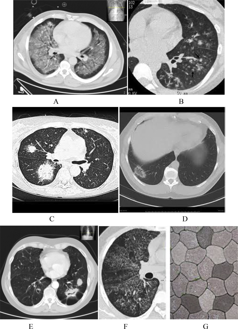
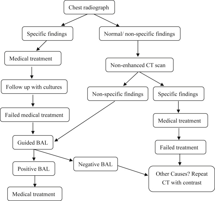
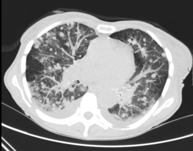
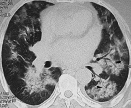
Respiratory infections and subsequent complications are one of the leading causes of high mortality in immunocompromised patients. Although chest radiograph and computed tomography are the commonly used diagnostic tools for the early diagnosis of lung manifestations of infections, they lack the specificity for the wide range of chest infections which can occur in immunocompromised patients. Systematic analysis of the imaging findings in correlation with the clinical settings along with comparison with the old images can expedite early and accurate diagnosis for subsequent appropriate management. Computer tomography findings in immunocompromised patients with respiratory infections, with regards to various clinical settings, will be discussed here.
Keywords: AIDS; Cancer drugs; Chest infection; Computed tomography; Immunocompromised patient; Pneumonia.
© 2016 Beijing You'an Hospital affiliated to Capital Medical University. Production and hosting by Elsevier B.V.
Figures
References
-
- Rubin R.H., Ferraro M.J. Understanding and diagnosing infectious complications in the immunocompromised host. Hematol Oncol Clin North Am. 1993;7:795–812. - PubMed
-
- Chanock S. Evolving risk factors for infectious complications of cancer therapy. Hematol Oncol Clin North Am. 1993;7:771–793. - PubMed
-
- Ketai L., Jordan K., Marom E.M. Imaging infection. Clin Chest Med. 2008;29:77–105. - PubMed
-
- Heussel C.P., Kauczor H.U., Heussel G., Fischer B., Mildenberger P., Thelen M. Early detection of pneumonia in febrile neutropenic patients: use of thin-section CT. AJR Am J Roentgenol. 1997;169:1347–1353. - PubMed
-
- Worthy S., Kang E.Y., Muller N.L. Acute lung disease in the immunocompromised host: differential diagnosis at high-resolution CT. Semin Ultrasound CT MR. 1995;16:353–360. - PubMed
Publication types
LinkOut - more resources
Full Text Sources