. 2020 Apr 17;5(16):9605-9613.
doi: 10.1021/acsomega.0c01038. eCollection 2020 Apr 28.

In Situ Absorption and Fluorescence Microspectroscopy Investigation of the Molecular Incorporation Process into Single Nanoporous Protein Crystals

Affiliations

In Situ Absorption and Fluorescence Microspectroscopy Investigation of the Molecular Incorporation Process into Single Nanoporous Protein Crystals

Takayuki Uwada et al. ACS Omega. .

Abstract

Protein crystals exhibit distinct three-dimensional structures, which contain well-ordered nanoporous solvent channels, providing a chemically heterogeneous environment. In this paper, the incorporation of various molecules into the solvent channels of native hen egg-white lysozyme crystals was demonstrated using fluorescent dyes, including acridine yellow G, rhodamine 6G, and eosin Y. The process was evaluated on the basis of absorption and fluorescence microspectroscopy at a single-crystal level. The molecular loading process was clearly visualized as a function of time, and it was determined that the protein crystals could act as nanoporous materials. It was found that the incorporation process is strongly dependent on the molecular charge, leading to heterogeneous molecular aggregation, which suggests host-guest interaction of protein crystals from the viewpoint of nanoporous materials.


Conflict of interest statement

The authors declare no competing financial interest.

Figures
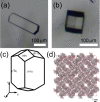
Figure 1
Figure 1
Transmittance microscopic images of hen egg-white lysozyme crystals obtained using the above experimental procedure. The (a) hexagonal and (b) square-shaped ones correspond to (110) and (101) faces of the tetragonal HEWL crystals, respectively. (c) Schematic illustration of the morphology of a tetragonal HEWL crystal, showing the crystallographic axes and faces. (d) c-axis projection of the structure of the tetragonal HEWL crystal.
Chart 1
Chart 1. Structures of Fluorescent Dye Molecules Incorporated in Hen Egg-White Lysozyme Crystals
Figure 2
Figure 2
Stereoscopic microscopic images of a single well of a 48-well crystallization plate containing already-grown hen egg-white lysozyme (HEWL) crystals with the mother solution immediately after injection of 400 μM eosin Y solution into the (a) well and (b) 1 day after the injection.
Figure 3
Figure 3
(a) Temporal change of transmittance and fluorescence images of a single HEWL crystal immediately after the injection of 400 μM acridine yellow G into the mother solution and up to 1 day following the injection. (b) Differential fluorescence image between the one obtained after 1 day and the one obtained immediately after the dye injection. (c) Temporal change of fluorescence intensity at inside (red) and outside (black) the crystal after the injection of acridine yellow G. The measured positions are indicated in image (a) (red and black crosses). (d) Space-resolved fluorescence spectra of the inside of a single HEWL crystal 1 day after acridine yellow G incorporation (red) and outside the crystal (black). The measured positions are indicated in image (a) (red and black crosses).
Figure 4
Figure 4
(a) Temporal changes of the transmittance and fluorescence images of a single HEWL crystal obtained immediately after the injection of 400 μM rhodamine 6G into the mother solution and up to 1 day. (b) Temporal changes of fluorescence intensities at inside (red) and outside (black) the crystal after the injection of rhodamine 6G. The measured positions are indicated in image (a) (red and black crosses). (c) Temporal changes of fluorescence intensity at inside (red) and outside (black) the crystal after the injection of rhodamine 6G. (c) Space-resolved absorption spectrum of the inside of a single HEWL crystal following rhodamine 6G incorporation (red). The spectrum was taken from the indicated position in image (a) (red cross). For comparison, an absorption spectrum of a 10 μM rhodamine 6G sodium acetate buffer solution is also shown (black). (d) Space-resolved fluorescence spectra of the inside of a single HEWL crystal 1 day after rhodamine 6G incorporation (red) and of the outside of the crystal (black). The measured positions are indicated in image (a) (red and black crosses).
Figure 5
Figure 5
(a) Temporal changes of the transmittance and fluorescence images of a single HEWL crystal obtained immediately after injection of 400 μM eosin Y into the mother solution and up to 1 day. (b) Temporal changes of fluorescence intensity at inside (blue) and outside (black) the crystal and the surface (red) after the injection of eosin Y. The measured positions are indicated in image (a) (blue, red, and black crosses). (c) Space-resolved absorption spectrum of the inside of a single HEWL crystal following eosin Y incorporation (red). The spectrum was taken from the position indicated in image (a) (blue and red crosses). For comparison, the absorption spectrum of a 10 μM eosin Y sodium acetate buffer solution is also shown (black). (d) Space-resolved fluorescence spectra of the inside and the surface of a single HEWL crystal 1 day after eosin Y incorporation (blue and red) and outside of the crystal (black). The measured positions are indicated in image (a) (blue, red, and black crosses).
Figure 6
Figure 6
Transmittance and fluorescence images of a single HEWL crystal, which directs the (101) face to the top side 1 day after injection of 400 μM eosin Y into the mother solution.
Figure 7
Figure 7
Schematic illustration of the molecular incorporation process into a HEWL crystal along the c-axis in the case of (a) cationic molecules (rhodamine 6G) and (b) anionic molecules (eosin Y).

Similar articles

Cited by

  • Production of Cross-Linked Lipase Crystals at a Preparative Scale.
    Fernández-Penas R, Verdugo-Escamilla C, Martínez-Rodríguez S, Gavira JA. Fernández-Penas R, et al. Cryst Growth Des. 2021 Mar 3;21(3):1698-1707. doi: 10.1021/acs.cgd.0c01608. Epub 2021 Feb 17. Cryst Growth Des. 2021. PMID: 34602865 Free PMC article.
  • Magnetite Mineralization inside Cross-Linked Protein Crystals.
    Savchenko M, Sebastian V, Lopez-Lopez MT, Rodriguez-Navarro A, Alvarez De Cienfuegos L, Jimenez-Lopez C, Gavira JA. Savchenko M, et al. Cryst Growth Des. 2023 Apr 28;23(6):4032-4040. doi: 10.1021/acs.cgd.2c01436. eCollection 2023 Jun 7. Cryst Growth Des. 2023. PMID: 37304398 Free PMC article.

References

    1. Abe S.; Maity B.; Ueno T. Design of a Confined Environment Using Protein Cages and Crystals for the Development of Biohybrid Materials. Chem. Commun. 2016, 52, 6496–6512. 10.1039/C6CC01355D. - DOI - PubMed
    1. Vilenchik L. Z.; Griffith J. P.; Clair N.; Navia M. A.; Margolin A. L. Protein Crystals as Novel Microporous Materials. J. Am. Chem. Soc. 1998, 120, 4290–4294. 10.1021/ja973449+. - DOI
    1. Margolin A. L.; Navia M. A. Protein Crystals as Novel Catalytic Materials. Angew. Chem., Int. Ed. 2001, 40, 2204–2222. 10.1002/1521-3773(20010618)40:12<2204::AID-ANIE2204>3.0.CO;2-J. - DOI - PubMed
    1. Brühwiler D.; Calzaferri G.; Torres T.; Ramm J. H.; Gartmann N.; Dieu L.-Q.; López-Duarte I.; Martínez-Díaz M. V. Nanochannels for Supramolecular Organization of Luminescent Guests. J. Mater. Chem. 2009, 19, 8040–8067. 10.1039/b907308f. - DOI
    1. Wan Y.; Zhao On the Controllable Soft-Templating Approach to Mesoporous Silicates. Chem. Rev. 2007, 107, 2821–2860. 10.1021/cr068020s. - DOI - PubMed