. 2019 Dec 31:14:1179069519889022.
doi: 10.1177/1179069519889022. eCollection 2019.

Gene Transfer in Rodent Nervous Tissue Following Hindlimb Intramuscular Delivery of Recombinant Adeno-Associated Virus Serotypes AAV2/6, AAV2/8, and AAV2/9

Affiliations

Gene Transfer in Rodent Nervous Tissue Following Hindlimb Intramuscular Delivery of Recombinant Adeno-Associated Virus Serotypes AAV2/6, AAV2/8, and AAV2/9

Asad Jan et al. Neurosci Insights. .

Abstract

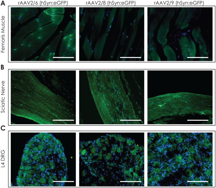
Recombinant adeno-associated virus (rAAV) vectors have emerged as the safe vehicles of choice for long-term gene transfer in mammalian nervous system. Recombinant adeno-associated virus-mediated localized gene transfer in adult nervous system following direct inoculation, that is, intracerebral or intrathecal, is well documented. However, recombinant adeno-associated virus delivery in defined neuronal populations in adult animals using less-invasive methods as well as avoiding ectopic gene expression following systemic inoculation remain challenging. Harnessing the capability of some recombinant adeno-associated virus serotypes for retrograde transduction may potentially address such limitations (Note: The term retrograde transduction in this manuscript refers to the uptake of injected recombinant adeno-associated virus particles at nerve terminals, retrograde transport, and subsequent transduction of nerve cell soma). In some studies, recombinant adeno-associated virus serotypes 2/6, 2/8, and 2/9 have been shown to exhibit transduction of connected neuroanatomical tracts in adult animals following lower limb intramuscular recombinant adeno-associated virus delivery in a pattern suggestive of retrograde transduction. However, an extensive side-by-side comparison of these serotypes following intramuscular delivery regarding tissue viral load, and the effect of promoter on transgene expression, has not been performed. Hence, we delivered recombinant adeno-associated virus serotypes 2/6, 2/8, or 2/9 encoding enhanced green fluorescent protein (eGFP), under the control of either cytomegalovirus (CMV) or human synapsin (hSyn) promoter, via a single unilateral hindlimb intramuscular injection in the bicep femoris of adult C57BL/6J mice. Four weeks post injection, we quantified viral load and transgene (enhanced green fluorescent protein) expression in muscle and related nervous tissues. Our data show that the select recombinant adeno-associated virus serotypes transduce sciatic nerve and groups of neurons in the dorsal root ganglia on the injected side, indicating that the intramuscular recombinant adeno-associated virus delivery is useful for achieving gene transfer in local neuroanatomical tracts. We also observed sparse recombinant adeno-associated virus viral delivery or eGFP transduction in lumbar spinal cord and a noticeable lack thereof in brain. Therefore, further improvements in recombinant adeno-associated virus design are warranted to achieve efficient widespread retrograde transduction following intramuscular and possibly other peripheral routes of delivery.

Keywords: Adeno-associated virus; gene delivery; nervous system; recombinant adeno-associated virus delivery.


Conflict of interest statement

Declaration of Conflicting Interests:The author(s) declared no potential conflicts of interest with respect to the research, authorship, and/or publication of this article.

Figures

Figure 1.
Figure 1.
Determination of vector genome copy numbers 4 weeks after a single right hindlimb intramuscular injection of rAAV particles in the injected (biceps femoris) muscle and other tissues. (A) Hindlimb biceps femoris muscle, (B) sciatic nerve, (C) L4-L5 dorsal root ganglia, and lumbar segment of (D) spinal cord, (E) brain, and (F) liver. (2/6, 2/8, and 2/9 indicate respective rAAV serotypes; n = 3/group, samples were run in duplicates; multiple group comparisons were assessed by one-way ANOVA post hoc Bonferroni test; pair-wise comparisons were further assessed by Mann-Whitney test; *P ⩽ .05, ***P ⩽ .005, only significant differences are highlighted in (A), (B), (C), and (F); NS in (D) and (E), not significant; error bars indicate mean ± s.e.m.). ANOVA indicates analysis of variance; CL, contralateral – uninjected side; CMV, cytomegalovirus promoter; hSyn, human synapsin promoter; IL, ipsilateral – injected side; rAAV, recombinant adeno-associated virus; s.e.m., standard error of mean.
Figure 2.
Figure 2.
RT-PCR to determine enhanced green fluorescent protein (eGFP) mRNA expression in the injected (biceps femoris) muscle and other tissues 4 weeks following unilateral hindlimb intramuscular delivery of rAAV2/6, 2/8, or 2/9 (CMV:eGFP). (A) Hindlimb biceps femoris muscle; (B) sciatic nerve; (C) L4-L5 dorsal root ganglia; and (D) liver, brain, and lumbar spinal cord segment. (eGFP mRNA relative ΔΔCT quantification was performed using mouse gapdh as the reference gene; data for rAAV2/6 are shown as black bars, data for rAAV2/8 are shown as grey bars, and data for rAAV2/9 are shown as white bars; n = 3/group, samples were run in duplicates; qPCR data in (D) were normalized to liver; multiple group comparisons were assessed by one-way ANOVA post hoc Bonferroni test; pair-wise comparisons were further assessed by Mann-Whitney test; *P ⩽ .05, **P ⩽ .01, and ***P ⩽ .005, only significant differences are highlighted in (A) to (C); NS in (D), not significant; error bars indicate mean ± s.e.m.). CL indicates contralateral – uninjected side; CMV, cytomegalovirus promoter; eGFP, enhanced green fluorescent protein; IL, ipsilateral – injected side; qPCR, quantitative PCR; rAAV, recombinant adeno-associated virus.
Figure 3.
Figure 3.
eGFP immunofluorescence in the injected (biceps femoris) muscle and other tissues on ipsilateral side 4 weeks following unilateral hindlimb intramuscular delivery of rAAV2/6, 2/8, or 2/9 (CMV:eGFP). Representative images showing eGFP immunofluorescence (green) in the (A) right hindlimb biceps femoris muscle, (B) right sciatic nerve, and (C) right L4 dorsal root ganglia (DRG) of mice injected with indicated rAAV serotypes. DAPI (blue) was used as a nuclear stain (20× magnified view; scale bar = 100 µm). Also see Figure S1 containing data for contralateral side (rAAV2/6-CMV:eGFP). CMV indicates cytomegalovirus promoter; DRG, dorsal root ganglia; eGFP, enhanced green fluorescent protein; rAAV, recombinant adeno-associated virus.
Figure 4.
Figure 4.
eGFP immunofluorescence in the lumbar spinal cord 4 weeks following unilateral hindlimb intramuscular delivery of rAAV2/6, 2/8, or 2/9 (CMV:eGFP). Representative images showing eGFP immunofluorescence (green) in lumbar spinal cord of mice injected with the rAAV serotypes (A) 2/6, (B) 2/8, or (C) 2/9. The sections were counterstained with NeuN (neuronal nuclei protein, in red) and DAPI nuclear stain (blue). The left panel shows panoramic image (5× magnification), and the corresponding high-magnification (20×) views in ventral horn lamina IX (VH) and dorsal horn lamina III-V (DH) are presented in the right side panel (scale bar = 100 μm). The insets show magnified view (80×) of neuronal processes (white box) and neuronal cell bodies (red box).
Figure 5.
Figure 5.
RT-PCR to determine enhanced green fluorescent protein (eGFP) mRNA expression in the injected (biceps femoris) muscle and other tissues 4 weeks following unilateral hindlimb intramuscular delivery of rAAV2/6, 2/8, or 2/9 (hSyn:eGFP). (A) Hindlimb biceps femoris muscle, (B) sciatic nerve, (C) L4-L5 dorsal root ganglia, and (D) liver, brain, and lumbar spinal cord segment (eGFP mRNA relative ΔΔCT quantification was performed using mouse gapdh as the reference gene; data for rAAV2/6 are shown as black bars, data for rAAV2/8 are shown as grey bars, and data for rAAV2/9 are shown as white bars; n = 3/group, samples were run in duplicates; qPCR data in (D) were normalized to liver; multiple group comparisons were assessed by one-way ANOVA post hoc Bonferroni test; pair-wise comparisons were further assessed by Mann-Whitney test; *P ⩽ .05, **P ⩽ .01, and ***P ⩽ .005, only significant differences are highlighted; NS in (B) and (D), not significant; error bars indicate mean ± s.e.m.). CL indicates contralateral uninjected side; eGFP, enhanced green fluorescent protein; hSyn, human synapsin promoter; IL, ipsilateral – injected side; qPCR, quantitative PCR; rAAV, recombinant adeno-associated virus.
Figure 6.
Figure 6.
eGFP immunofluorescence in the injected (biceps femoris) muscle and other tissues on ipsilateral side 4 weeks following unilateral hindlimb intramuscular delivery of rAAV2/6, 2/8, or 2/9 (hSyn:eGFP). Representative images showing eGFP immunofluorescence (green) in the (A) right hindlimb biceps femoris muscle, (B) right sciatic nerve, and (C) right L4 dorsal root ganglia (DRG) of mice injected with indicated rAAV serotypes. DAPI (blue) was used as a nuclear stain; 20× magnified view; scale bar = 100 µm. Also see Figure S1 containing data for contralateral side (rAAV2/6-hSyn:eGFP). DRG indicates dorsal root ganglia; eGFP, enhanced green fluorescent protein; hSyn, human synapsin promoter; rAAV, recombinant adeno-associated virus.
Figure 7.
Figure 7.
eGFP immunofluorescence in the lumbar spinal cord 4 weeks following unilateral hindlimb intramuscular delivery of rAAV2/6, 2/8, or 2/9 (hSyn:eGFP). Representative images showing eGFP immunofluorescence (green) in lumbar spinal cord of mice injected with the rAAV serotypes 2/6 (A), 2/8 (B), or 2/9 (C). The sections were counterstained with NeuN (neuronal nuclei protein, in red) and DAPI nuclear stain (blue). The left panel shows panoramic image (5× magnification), and the corresponding high-magnification (20×) views in ventral horn lamina IX (VH) and dorsal horn lamina III-V (DH) are presented in the right side panel (scale bar = 100 µm). The insets show magnified view (80×) of neuronal processes (white box) and neuronal cell bodies (red box). DH indicates dorsal horn; eGFP, enhanced green fluorescent protein; hSyn, human synapsin promoter; rAAV, recombinant adeno-associated virus; VH, ventral horn.

Similar articles

Cited by

References

    1. Bedbrook CN, Deverman BE, Gradinaru V. Viral strategies for targeting the central and peripheral nervous systems. Annu Rev Neurosci. 2018;41:323-348. - PubMed
    1. Calcedo R, Vandenberghe LH, Gao G, Lin J, Wilson JM. Worldwide epidemiology of neutralizing antibodies to adeno-associated viruses. J Infect Dis. 2009;199:381-390. - PMC - PubMed
    1. Gao G, Vandenberghe LH, Alvira MR, et al. Clades of adeno-associated viruses are widely disseminated in human tissues. J Virol. 2004;78:6381-6388. - PMC - PubMed
    1. Hudry E, Vandenberghe LH. Therapeutic AAV gene transfer to the nervous system: a clinical reality. Neuron. 2019;101:839-862. - PMC - PubMed
    1. Lykken EA, Shyng C, Edwards RJ, Rozenberg A, Gray SJ. Recent progress and considerations for AAV gene therapies targeting the central nervous system. J Neurodev Disord. 2018;10:16. - PMC - PubMed

LinkOut - more resources