Comparative Study
. 2020 Jul 1;15(5):511-522.
doi: 10.1093/scan/nsaa060.

Comparing two facets of emotion perception across multiple neurodegenerative diseases

Affiliations
Comparative Study

Comparing two facets of emotion perception across multiple neurodegenerative diseases

Casey L Brown et al. Soc Cogn Affect Neurosci. .

Abstract

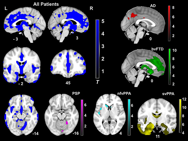
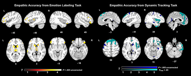
Deficits in emotion perception (the ability to infer others' emotions accurately) can occur as a result of neurodegeneration. It remains unclear how different neurodegenerative diseases affect different forms of emotion perception. The present study compares performance on a dynamic tracking task of emotion perception (where participants track the changing valence of a film character's emotions) with performance on an emotion category labeling task (where participants label specific emotions portrayed by film characters) across seven diagnostic groups (N = 178) including Alzheimer's disease (AD), behavioral variant frontotemporal dementia (bvFTD), semantic variant primary progressive aphasia (svPPA), non-fluent variant primary progressive aphasia (nfvPPA), progressive supranuclear palsy (PSP), corticobasal syndrome and healthy controls. Consistent with hypotheses, compared to controls, the bvFTD group was impaired on both tasks. The svPPA group was impaired on the emotion labeling task, whereas the nfvPPA, PSP and AD groups were impaired on the dynamic tracking task. Smaller volumes in bilateral frontal and left insular regions were associated with worse labeling, whereas smaller volumes in bilateral medial frontal, temporal and right insular regions were associated with worse tracking. Findings suggest labeling and tracking facets of emotion perception are differentially affected across neurodegenerative diseases due to their unique neuroanatomical correlates.

Keywords: cognitive empathy; dementia; emotion recognition; empathic accuracy; lesion.


Figures

Fig. 1
Fig. 1
Emotion perception task performance by diagnostic group. Error bars represent 95% confidence intervals. Blue asterisks reflect differences on the emotion category labeling task between group, red asterisks reflect differences on the dynamic valence tracking task and black asterisks reflect within group differences.
Fig. 2
Fig. 2
T-score maps of brain areas for which volume loss was greater than HCs, when controlling for age, gender, handedness, total intracranial volume and scanner field strength (PFWE < 0.05). Compared to controls, the patient group had smaller volume in the frontal (e.g. medial prefrontal and orbitofrontal cortex, anterior cingulate cortex and insula), striatum and temporal (e.g. caudate, putamen, amygdala and hippocampus) and parietal regions (e.g. precuneus). Thus, patients altogether showed a widespread pattern of neurodegeneration. The CBS group did not show significant volume loss compared to controls at this threshold.
Fig. 3
Fig. 3
T-score maps of brain areas for which volume loss was associated with worse emotion perception on the emotion labeling film task and dynamic tracking task, when controlling for diagnosis, age, gender, handedness, disease severity, cognitive functioning, total intracranial volume and scanner field strength. Smaller volume (maximum T-score = 4.16) in bilateral inferior frontal, bilateral caudate, bilateral thalamus and left insula regions was associated with worse emotion labeling (P < 0.005, uncorrected). Smaller volume (maximum T-score = 5.22) in bilateral and predominantly right medial frontal, right anterior and middle temporal, right insular and bilateral cingulate and pre- and post-central gyri regions was associated with worse emotion perception (P < 0.005, uncorrected). Two large clusters in superior medial frontal and right anterior temporal and insular areas were also significant with family wise error correction (PFWE < 0.05) for the dynamic tracking task.

References

    1. Abdulkadir A., Mortamet B., Vemuri P., Jack C.R., Krueger G., Klöppel S. (2011). Effects of hardware heterogeneity on the performance of SVM Alzheimer’s disease classifier. NeuroImage, 58(3), 785–92. - PMC - PubMed
    1. Allender J., Kaszniak A.W. (1989). Processing of emotional cues in patients with dementia of the Alzheimer’s type. The International Journal of Neuroscience, 46(3–4), 147–55. - PubMed
    1. Armstrong M.J., Litvan I., Lang A.E., Bak T.H., Bhatia K.P., Borroni B., et al. (2013). Criteria for the diagnosis of corticobasal degeneration. Neurology, 80(5), 496–503. - PMC - PubMed
    1. Ashburner J., Friston K.J. (2005). Unified segmentation. NeuroImage, 26(3), 839–51. - PubMed
    1. Baez S., Manes F., Huepe D., Torralva T., Fiorentino N., Richter F., et al. (2014). Primary empathy deficits in frontotemporal dementia. Frontiers in Aging Neuroscience, 6, 262. - PMC - PubMed

Publication types