. 2020 Dec 9;30(12):981-988.
doi: 10.1093/glycob/cwaa042.

Deducing the N- and O-glycosylation profile of the spike protein of novel coronavirus SARS-CoV-2

Affiliations

Deducing the N- and O-glycosylation profile of the spike protein of novel coronavirus SARS-CoV-2

Asif Shajahan et al. Glycobiology. .

Abstract

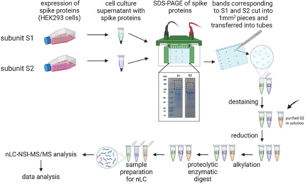
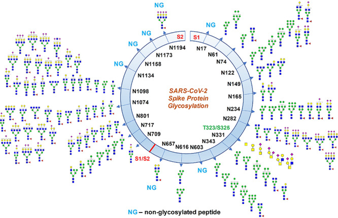
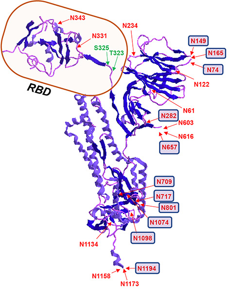
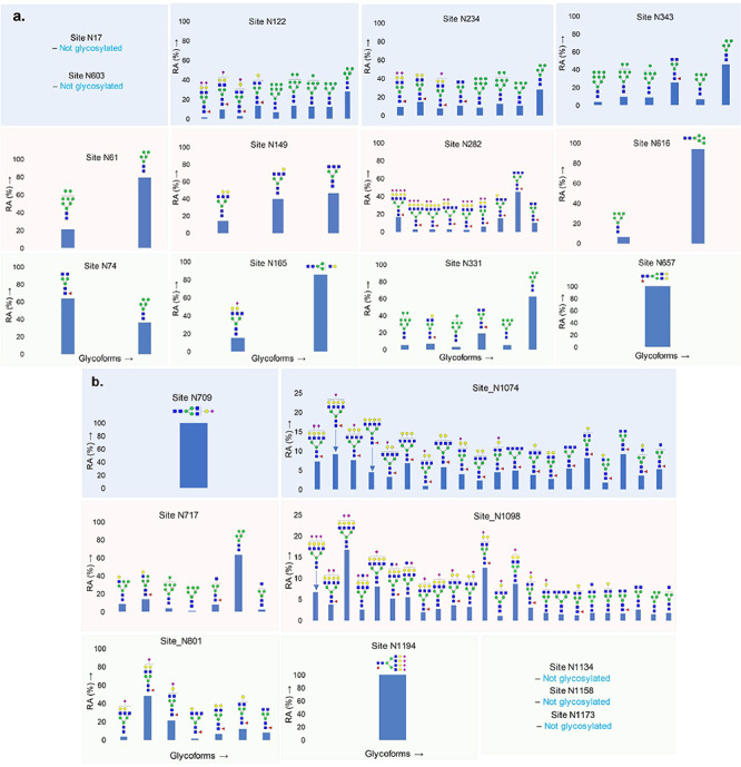
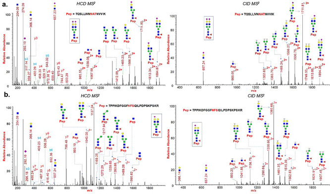
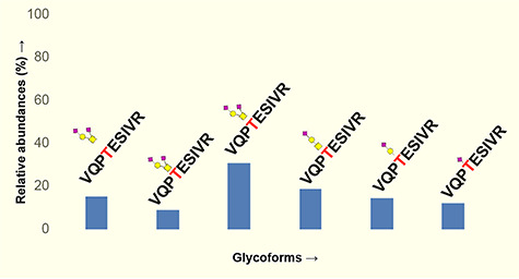
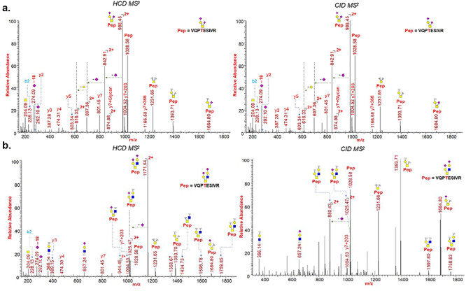
The current emergence of the novel coronavirus pandemic caused by severe acute respiratory syndrome coronavirus 2 (SARS-CoV-2) demands the development of new therapeutic strategies to prevent rapid progress of mortalities. The coronavirus spike (S) protein, which facilitates viral attachment, entry and membrane fusion is heavily glycosylated and plays a critical role in the elicitation of the host immune response. The spike protein is comprised of two protein subunits (S1 and S2), which together possess 22 potential N-glycosylation sites. Herein, we report the glycosylation mapping on spike protein subunits S1 and S2 expressed on human cells through high-resolution mass spectrometry. We have characterized the quantitative N-glycosylation profile on spike protein and interestingly, observed unexpected O-glycosylation modifications on the receptor-binding domain of spike protein subunit S1. Even though O-glycosylation has been predicted on the spike protein of SARS-CoV-2, this is the first report of experimental data for both the site of O-glycosylation and identity of the O-glycans attached on the subunit S1. Our data on the N- and O-glycosylation are strengthened by extensive manual interpretation of each glycopeptide spectra in addition to using bioinformatics tools to confirm the complexity of glycosylation in the spike protein. The elucidation of the glycan repertoire on the spike protein provides insights into the viral binding studies and more importantly, propels research toward the development of a suitable vaccine candidate.

Keywords: COVID-19; S1 S2 glycosylation; SARS-CoV-2 glycosylation; coronavirus vaccine; spike protein.


Figures

Fig. 1
Fig. 1
The SARS-CoV-2 spike proteins recombinantly expressed on HEK293 supernatant were fractionated through SDS-PAGE, subsequently digested by proteases and analyzed by nLC-NSI-MS/MS. The expression of SARS-CoV-2 spike protein subunits S1 and S2 on HEK 293 culture supernatant showed higher molecular weight upon SDS-PAGE than expected, because of glycosylation. Thus, the gel bands corresponding to the molecular weight of 200–100 kDa for S1 and 150–80 kDa for S2 were cut, proteins were lysed after reduction–alkylation and analyzed by LC-MS/MS (created with biorender.com). Purified S2 were processed after in-solution protease digestion.
Fig. 2
Fig. 2
Glycosylation profile on coronavirus SARS-CoV-2 characterized by high-resolution LC-MS/MS. About 17 N-glycosylation sites were found occupied out of 22 potential sites along with two O-glycosylation sites bearing core-1 type O-glycans. Some N-glycosylation sites were partially glycosylated. Monosaccharide symbols follow the Symbol Nomenclature for Glycans (SNFG) system (Varki et al. 2015).
Fig. 3
Fig. 3
3D structure of SARS-CoV-2 spike glycoprotein showing the location of N- and O-glycosylation. Receptor-binding domain (RBD) is highlighted and sites with predominant complex type N-glycans are represented with small squares. PBD ID: 6VXX: only one monomer is shown after removing the ligands.
Fig. 4
Fig. 4
Quantitative glycosylation profile of N-glycans on coronavirus SARS-CoV-2 spike protein characterized by high-resolution LC-MS/MS. (A) 13 sites on subunit S1; (B) 9 sites on subunit S2. RA: relative abundances. Monosaccharide symbols follow the SNFG system (Varki et al. 2015).
Fig. 5
Fig. 5
HCD and CID MS/MS spectra showing glycan neutral losses, oxonium ions and peptide fragments of (A) representative N-glycopeptide TQSLLIVNNATNVVIK (site N122) of spike protein subunit S1; (B) representative N-glycopeptide TPPIKDFGGFNFSQILPDPSKPSKR (site N801) of spike protein subunit S2. Monosaccharide symbols follow the SNFG system (Varki et al. 2015).
Fig. 6
Fig. 6
Quantitative glycosylation profile of O-glycans on sites T323 of coronavirus SARS-CoV-2 spike protein characterized by high-resolution LC-MS/MS. Monosaccharide symbols follow the SNFG system (Varki et al. 2015).
Fig. 7
Fig. 7
HCD and CID MS/MS spectra showing glycan neutral losses, oxonium ions and peptide fragments of (A) representative O-Glycopeptide 320VQPTESIVR328 with core 1 type GalNAcGalNeuAc2 glycan detected on site Thr323 of spike protein subunit S1; (B) representative O-Glycopeptide 320VQPTESIVR328 with core 2 type GalNAcGalNeuAc(GlcNAcGalNeuAc) glycan detected on site Thr323 of spike protein subunit S1. Monosaccharide symbols follow the SNFG system (Varki et al. 2015).

References

    1. Afrough B, Dowall S, Hewson R. 2019. Emerging viruses and current strategies for vaccine intervention. Clin Exp Immunol. 196:157–166. - PMC - PubMed
    1. Andersen KG, Rambaut A, Lipkin WI, Holmes EC, Garry RF. 2020. The proximal origin of SARS-CoV-2. Nat Med 26:450–452. - PMC - PubMed
    1. Bagdonaite I, Wandall HH. 2018. Global aspects of viral glycosylation. Glycobiology. 28:443–467. - PMC - PubMed
    1. Chakraborti S, Prabakaran P, Xiao X, Dimitrov DS. 2005. The SARS coronavirus S glycoprotein receptor binding domain: Fine mapping and functional characterization. Virol J. 2:73. - PMC - PubMed
    1. Chang D, Zaia J. 2019. Why glycosylation matters in building a better flu vaccine. Mol Cell Proteomics. 18:2348–2358. - PMC - PubMed

Publication types

Substances