

123I-FP-CIT SPECT validation of nigro-putaminal MRI tractography in dementia with Lewy bodies
- PMID: 32363488
- PMCID: PMC7196565
- DOI: 10.1186/s41747-020-00153-6
123I-FP-CIT SPECT validation of nigro-putaminal MRI tractography in dementia with Lewy bodies
Abstract
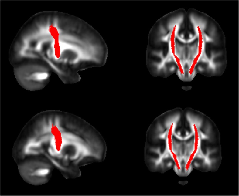
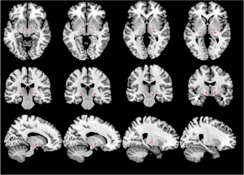
Background: Assessment of nigrostriatal degeneration is a key element to discriminate between dementia with Lewy bodies (DLB) and Alzheimer disease (AD), and it is often evaluated using ioflupane (123I-FP-CIT) single-photon emission computed tomography (SPECT). Given the limited availability of 123I-FP-CIT SPECT, we evaluated if a mask-based approach to nigroputaminal magnetic resonance imaging (MRI) diffusion-weighted tractography could be able to capture microstructural changes reflecting nigroputaminal degeneration in DLB.
Methods: A nigroputaminal bundle mask was delineated on 12 healthy volunteers (HV) and applied to MRI diffusion-weighted data of 18 subjects with DLB, 21 subjects with AD and another group of 12 HV. The correlation between nigroputaminal fractional anisotropy (FA) values and 123I-FP-CIT SPECT findings was investigated. Shapiro-Wilk, ANOVA, ANCOVA, and parametric correlation statistics as well as receiver operating characteristic (ROC) analysis were used.
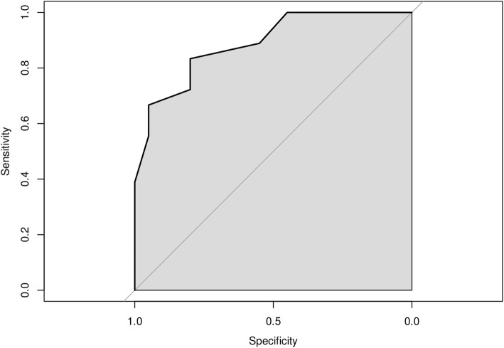
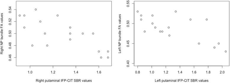
Results: DLB patients showed a higher nigroputaminal FA values compared with both AD and HV-controls groups (p = 0.001 for both comparisons), while no difference was observed between HV-controls and AD groups (p = 0.450); at ROC analysis, the area under the curve for the discriminating DLB and AD subjects was 0.820; FA values correlated with 123I-FP-CIT values (on the left, r = -0.670; on the right, r = -720). No significant differences were observed for the FA of the corticospinal tract across the three groups (p = 0.740).
Conclusions: In DLB, nigroputaminal degeneration could be reliably assessed on MRI diffusion scans using a mask of nigroputaminal bundle trajectory. Nigroputaminal FA in DLB patients correlated with 123I-FP-CIT values data may allow to differentiate these patients from AD patients and HV-controls.
Keywords: Alzheimer disease; Diffusion tensor imaging; Ioflupane; Magnetic resonance imaging; Tomography (emission-computed; single-photon).
Conflict of interest statement
MP receives research support from Novartis and received honoraria from Merck and Novartis.
FN has received fees for board participation by Roche and Eli-Lilly, and speaker honoraria from Eli-Lilly.
SM has received speaker honoraria from GE healthcare and Eli Lilly.
GLM has received honoraria for lecturing, travel expenses for attending meetings and financial support for research from Bayer Schering, Biogen Idec, Sanofi Aventis, Teva, Genzyme, and Merck Serono Pharmaceuticals.
The other authors declare that they have no competing interests.
Figures
References
-
- McKeith I, Mintzer J, Aarsland D et al (2004) Dementia with Lewy bodies. Lancet Neurol 3:19– 28. 10.1016/s1474-4422(03)00619-7 - PubMed