Review
. 2021 Mar;52(1):125-134.
doi: 10.1007/s42770-020-00274-5. Epub 2020 May 3.

Feline sporotrichosis in Asia

Affiliations
Review

Feline sporotrichosis in Asia

Hock Siew Han et al. Braz J Microbiol. 2021 Mar.

Abstract

Sporothrix schenckii sensu lato is currently recognized as a species complex with only Sporothrix brasiliensis, Sporothrix schenckii sensu stricto, Sporothrix globosa and Sporothrix pallida identified to cause disease in the cat. Feline sporotrichosis in Asia is mainly reported from Malaysia where a single clonal strain of clinical clade D, Sporothrix schenckii sensu stricto manifesting low susceptibility to major antifungal classes, has been identified as the agent of the disease. Sporothrix globosa has been identified to cause disease from a single cat in Japan while the specific species of agent has not been identified yet for the disease in Thailand. Despite efforts to elucidate and describe the pathogenicity of the agent and the disease it causes, the paucity of data highlights the need for further molecular epidemiological studies to characterize this fungus and the disease it causes in Asia. Its prognosis remains guarded to poor due to issues pertaining to cost, protracted treatment course, zoonotic potential and low susceptibility of some strains to antifungals.

Keywords: Asia; Feline sporotrichosis; Malaysia.


Conflict of interest statement

The authors declare that there are no conflict of interest.

Figures

Fig. 1
Fig. 1
A solitary chronic, non-healing and exudative lesion at the paw of a cat with sporotrichosis
Fig. 2
Fig. 2
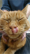
Chronic, non-healing ulcers on the bridge of the nose by S. schenckii sensu stricto infection of a cat in Malaysia. Note that small granulomas are forming at the tip of both ears
Fig. 3
Fig. 3
As the disease progresses, non-encapsulated granulomas form that then ulcerate at ear tips. These exudate then dry to form crusts
Fig. 4
Fig. 4
Some cats are presented with generalized, chronic, non-healing nodular lesions at the thorax extending to the abdomen, not just at the head and ear tips
Fig. 5
Fig. 5
A dorsal view of a cat with pyogranulomatous nodular and exudative lesions at the ear tips, highlighting the preponderance of the fungus to aggregate at the cooler regions of the host
Fig. 6
Fig. 6
Colony morphology of S. schenckii sensu stricto. Black mycelial form colony was observed on Sabouraud glucose agar after culture at 28 °C for 14 days (a). White yeast-like colony was observed on Sabouraud glucose agar after culture at 37 °C for 10 days (b)
Fig. 7
Fig. 7
Yeasts are found in abundance from direct impression smears from the feline patient. They are located intra and extracellularly, in pleomorphic shapes ranging from cigar-shaped to round or oval, measuring 3–5 μm in diameter with a thin, clear halo around a pale blue cytoplasm. Haemacolor stain
Fig. 8
Fig. 8
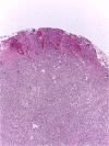
Diffuse pyogranulomatous inflammation is seen throughout the superficial and deep dermis that may extend to the deeper subcutis
Fig. 9
Fig. 9
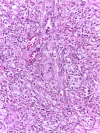
Round to cigar-shaped organism, seen both free and within macrophages. Commonly, the yeast in cytoplasm of macrophages creates large clear pockets full of yeast due to poorly visualized yeast cell wall
Fig. 10
Fig. 10
A cat with Cryptococcosis. The involvement of the nasal sinuses and occasional cutaneous spread of the disease often confuses the veterinarian between cryptococcosis with that of sporotrichosis
Fig. 11
Fig. 11
A cat with squamous cell carcinoma (SCC) confirmed via histopathology. As the most common form of malignant neoplasm of the feline skin, SCC is associated with sun-damaged skin with the nasal planum, pinnae and eyelids the most common affected sites. Its clinical predisposition site and appearance may be easily confused with that of sporotrichosis
Fig. 12
Fig. 12
A cat with herpesvirus-1 (feline rhinotracheitis) infection. There is extensive ulcerative and necrotizing dermatitis involving the nasal planum, bridge of nose and periocular skin with concurrent respiratory signs. Very similar with those of a cat with sporotrichosis with lesion extension to the nasal sinus which causes secondary respiratory dyspnoea. Image courtesy of Dr. Francesco Albanese
Fig. 13
Fig. 13
A cat with an erosive and ulcerative nodular lesion at the left peri-nasal orifice caused by Mycobacterium microti which is clinically very similar with sporotrichosis. Image courtesy of Prof. Danielle Gunn-Moore
Fig. 14
Fig. 14
Post-healing ear tip deformity due to extensive tissue necrosis from infection

Similar articles

Cited by

References

    1. Zhou X, Rodrigues A, Feng P, Hoog GS. Global ITS diversity in the Sporothrix schenckii complex. Fungal Divers. 2013;2013:1–13.
    1. Rodrigues AM, Teixeira MM, Hoog GS, et al. Phylogenetic analysis reveals a high prevalence of Sporothrix brasiliensis in feline Sporotrichosis outbreaks. PLoS Negl Trop Dis. 2013;7:e2281. - PMC - PubMed
    1. Han HS, Kano R, Chen C, Noli C. Comparisons of two in vitro antifungal sensitivity tests and monitoring during therapy of Sporothrix schenckii sensu stricto in Malaysian cats. Vet Dermatol. 2017;28:156–e32. - PubMed
    1. Kano R, Tsui CKM, Hamelin RC, Anzawa K, Mochizuki T, Nishimoto K, Hiruma M, Kamata H, Hasegawa A. The MAT1-1:MAT1-2 ratio of Sporothrix globosa isolates in Japan. Mycopathologia. 2015;179:81–86. - PubMed
    1. Schubach TMP, Menezes RC, Wanke B. Sporotrichosis. In: Greene CE, editor. Infectious diseases of the dog and cat. 4. St. Louis: Saunders Elsevier; 2012. pp. 645–650.

Substances

LinkOut - more resources