. 2020 Jul;13(4):1228-1244.
doi: 10.1111/1751-7915.13583. Epub 2020 May 3.

Pediococcus pentosaceus LI05 alleviates DSS-induced colitis by modulating immunological profiles, the gut microbiota and short-chain fatty acid levels in a mouse model

Affiliations

Pediococcus pentosaceus LI05 alleviates DSS-induced colitis by modulating immunological profiles, the gut microbiota and short-chain fatty acid levels in a mouse model

Xiaoyuan Bian et al. Microb Biotechnol. 2020 Jul.

Abstract

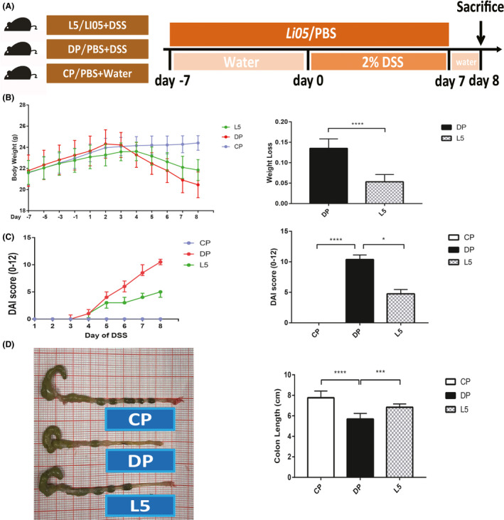
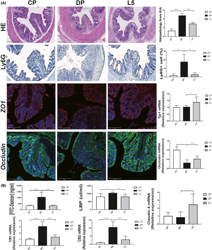
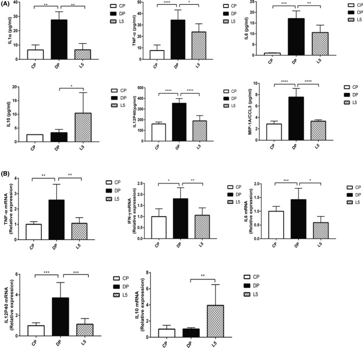
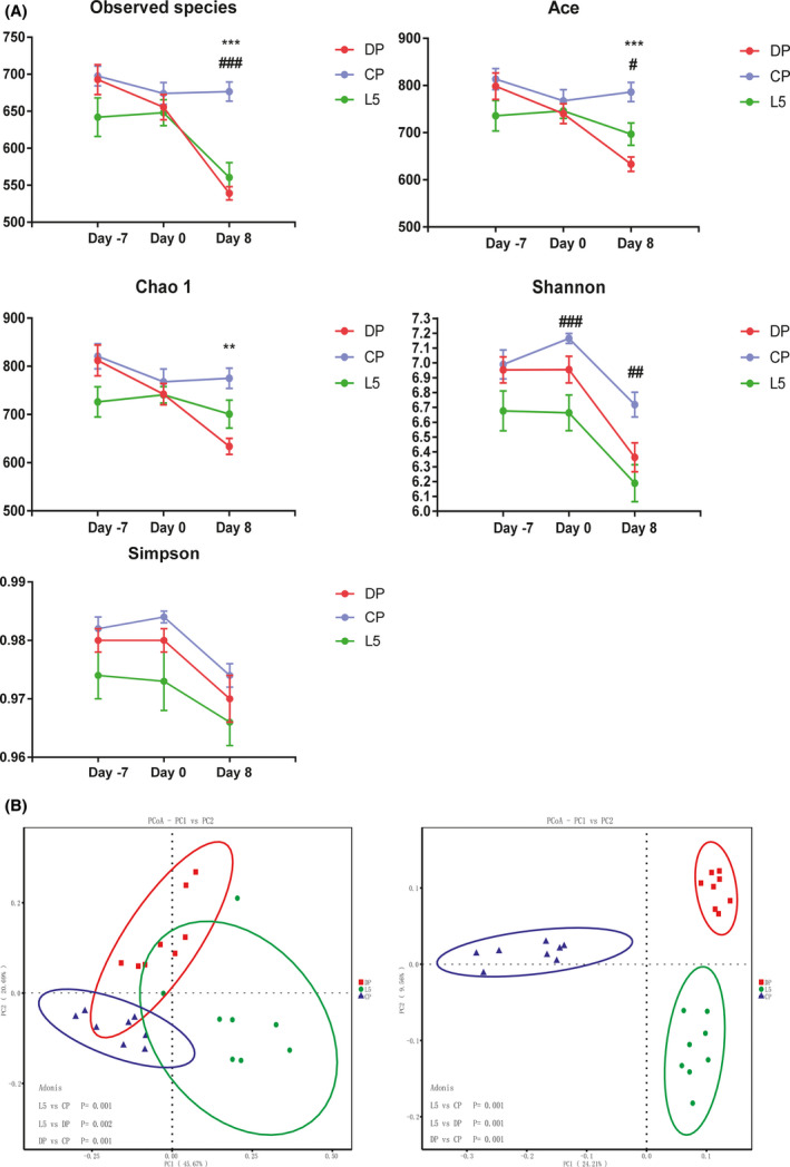
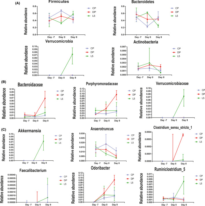
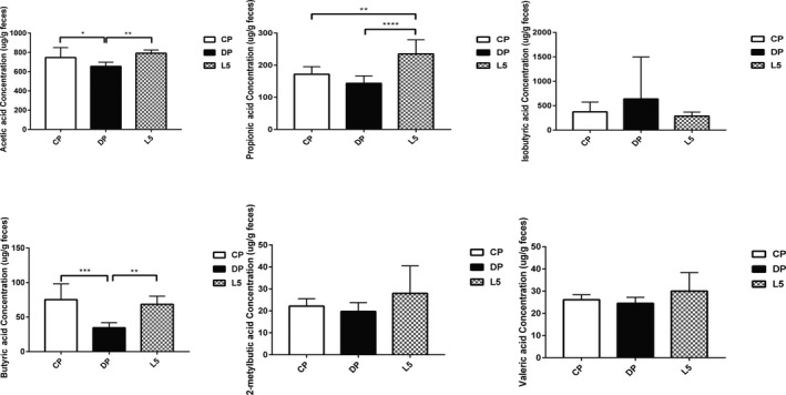
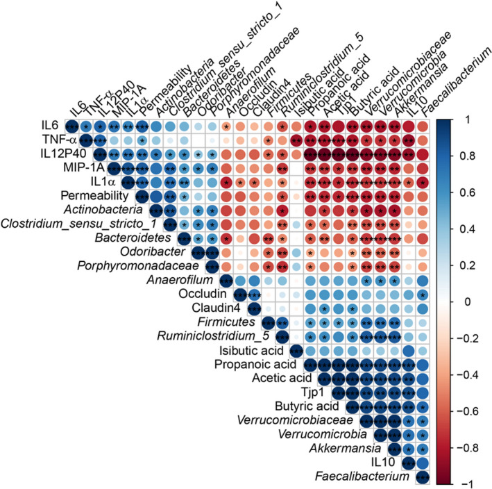
The gut microbiota is considered a key factor in pathogenesis and progression of inflammatory bowel disease (IBD). The bacterium Pediococcus pentosaceus LI05 alleviated host inflammation by maintaining the gut epithelial integrity, modulating the host immunity, gut microbiota and metabolism, but its effect on IBD remains unclear. The present study aimed to investigate the role and mechanisms of P. pentosaceus LI05. Mice were administered P. pentosaceus LI05 or phosphate-buffered saline once daily by oral gavage for 14 days, and colitis was induced by providing mice 2% DSS-containing drinking water for 7 days. P. pentosaceus LI05 ameliorated colitis in mice and reduced the body weight loss, disease activity index (DAI) scores, colon length shortening, intestinal permeability and the proinflammatory cytokine levels. Furthermore, a significantly altered gut microbiota composition with increased diversity and short-chain fatty acid (SCFA) production was observed in mice treated with P. pentosaceus LI05. Several genera, including Akkermansia and Faecalibacterium, were differentially enriched in the P. pentosaceus LI05-treated mice and were negatively correlated with colitis indices and positively correlated with gut barrier markers and SCFA levels. The P. pentosaceus LI05 treatment alleviated intestinal inflammation by maintaining the intestinal epithelial integrity and modulating the immunological profiles, gut microbiome and metabolite composition. Based on our findings, P. pentosaceus LI05 might be applied as potential preparation to ameliorate colitis.


Conflict of interest statement

The authors declare no conflicts of interest.

Figures

Fig. 1
Fig. 1
P. pentosaceus LI05 ameliorated DSS‐induced clinical symptoms and intestinal injury in mice. A. Schematic depicting the design of the animal experiment. B. The body weights of mice in the three groups recorded from day −7 to day 8 are plotted and presented as mean (SD; left panel). The bar chart represents the weight loss relative to the base weight on day 0 (right panel). C. The disease activity index (DAI) scores after DSS administration (left panel) and on day 8 (right panel) are shown for the three groups. D. Representative images of the colon in mice from the three groups (left panel) and the colon length on day 8 (right panel). *P < 0.05, ***P < 0.001 and ****P < 0.0001 according to unpaired t tests with Welch’s correction, Kruskal–Wallis tests and post hoc one‐way ANOVA.
Fig. 2
Fig. 2
P. pentosaceus LI05 ameliorated colon epithelial damage. A. Representative images of H&E staining, Ly6G immunohistochemical staining, ZO1 and Occludin immunofluorescence staining among three groups (left panel). Histopathology scores, the percentage of Ly6G‐positive cells, and colon expression of ZO1 (Tjp1) and Occludin in the three groups (right panel). B. The bar chart presents the plasma concentration of FITC–dextran, serum LBP levels and colon expression of CB1, CB2 and Claudin 4 among three groups. All data are presented as means ± SEM. *P < 0.05, **P < 0.01, ****P < 0.0001 according to the post hoc one‐way ANOVA.
Fig. 3
Fig. 3
P. pentosaceus LI05 ameliorated DSS‐induced colitis by exerting anti‐inflammatory effects. A. The serum levels of the cytokines IL1α, TNF‐α, IL6, IL10, IL12P40 and MIP‐1A in the three groups. B. Colon expression of TNF‐α, IFN‐γ, IL6, IL12P40 and IL10 in the three groups. All data are presented as means ± SEM. *P < 0.05, **P < 0.01, ***P < 0.001 and ****P < 0.0001 according to the post hoc one‐way ANOVA.
Fig. 4
Fig. 4
P. pentosaceus LI05 ameliorated gut microbiome diversity induced by DSS administration. A. Richness and diversity of the faecal microbiota in the three groups. Abbreviation: *CP group compared to the DP group. #CP group compared to the L5 group. All data are presented as means ± SEM. B. PCoA plot based on the weighted (left panel) and unweighted (right panel) UniFrac distances among the three groups (day 8). Each point represents a sample.
Fig. 5
Fig. 5
Dynamic changes in the abundance of specific taxa at the phylum (A), family (B) and genus (C) levels among the three groups during the experiment. All data are presented as medians with interquartile ranges.
Fig. 6
Fig. 6
P. pentosaceus LI05 administration increased the production of short‐chain fatty acids (SCFAs). The faecal concentration of SCFAs was determined using GC‐MS, including acetic acid, propionic acid, isobutyric acid, butyric acid, 2‐metylbutic acid and valeric acid. All data are presented as means ± SEM. *P < 0.05, **P < 0.01, ***P < 0.001 and ****P < 0.0001 according to the post hoc one‐way ANOVA.
Fig. 7
Fig. 7
Correlations of representative microbial taxa, gut barrier markers, inflammatory cytokines and SCFAs between the DP and L5 groups. Spearman’s rank correlation coefficients were calculated. Blue indicates positive correlations, and red indicates negative correlations. Significant differences were indicated as follows: *P < 0.05, **P < 0.01 and ***P < 0.001.

Similar articles

Cited by

References

    1. Ahl, D. , Liu, H. , Schreiber, O. , Roos, S. , Phillipson, M. , and Holm, L. (2016) Lactobacillus reuteri increases mucus thickness and ameliorates dextran sulphate sodium‐induced colitis in mice. Acta Physiol (Oxf) 217: 300–310. - PubMed
    1. Arpaia, N. , Campbell, C. , Fan, X. , Dikiy, S. , van der Veeken, J. , deRoos, P. , et al (2013) Metabolites produced by commensal bacteria promote peripheral regulatory T‐cell generation. Nature 504: 451–455. - PMC - PubMed
    1. Atarashi, K. , Tanoue, T. , Oshima, K. , Suda, W. , Nagano, Y. , Nishikawa, H. , et al (2013) Treg induction by a rationally selected mixture of Clostridia strains from the human microbiota. Nature 500: 232–236. - PubMed
    1. Bengmark, S. , Di Cocco, P. , Clemente, K. , Corona, L. , Angelico, R. , and Manzia, T. , et al (2011) Bio‐ecological control of chronic liver disease and encephalopathy. Minerva Med 102: 309–319. - PubMed
    1. Bian, X. , Wu, W. , Yang, L. , Lv, L. , Wang, Q. , Li, Y. , et al (2019) Administration of Akkermansia muciniphila Ameliorates Dextran sulfate sodium‐induced ulcerative colitis in mice. Front Microbiol 10: 2259. - PMC - PubMed

Publication types