. 2020 May 4;16(5):e1008244.
doi: 10.1371/journal.ppat.1008244. eCollection 2020 May.

Tuning antiviral CD8 T-cell response via proline-altered peptide ligand vaccination

Affiliations

Tuning antiviral CD8 T-cell response via proline-altered peptide ligand vaccination

Adil Doganay Duru et al. PLoS Pathog. .

Abstract

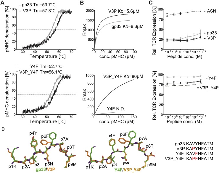
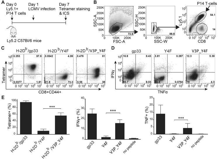
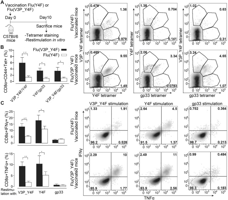
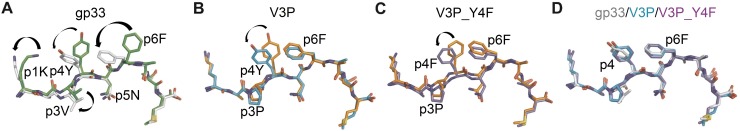
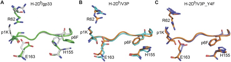
Viral escape from CD8+ cytotoxic T lymphocyte responses correlates with disease progression and represents a significant challenge for vaccination. Here, we demonstrate that CD8+ T cell recognition of the naturally occurring MHC-I-restricted LCMV-associated immune escape variant Y4F is restored following vaccination with a proline-altered peptide ligand (APL). The APL increases MHC/peptide (pMHC) complex stability, rigidifies the peptide and facilitates T cell receptor (TCR) recognition through reduced entropy costs. Structural analyses of pMHC complexes before and after TCR binding, combined with biophysical analyses, revealed that although the TCR binds similarly to all complexes, the p3P modification alters the conformations of a very limited amount of specific MHC and peptide residues, facilitating efficient TCR recognition. This approach can be easily introduced in peptides restricted to other MHC alleles, and can be combined with currently available and future vaccination protocols in order to prevent viral immune escape.


Conflict of interest statement

The authors have declared that no competing interests exist.

Figures

Fig 1
Fig 1. The p3P modification enhances pMHC stability without altering structural conformation, reestablishing TCR recognition.
A. The p3P modification increases pMHC stability. CD melting curves of H-2Db/gp33 and H-2Db/V3P (upper panel), and H-2Db/Y4F and H-2Db/V3P_Y4F (lower panel). Melting temperatures (Tm) corresponding to 50% protein denaturation are indicated. B. The soluble TCR P14 binds to the APL V3P_Y4F. In contrast to Y4F, V3P_Y4F is bound by P14. Binding affinity of the soluble TCR P14 to each pMHC was measured using SPR. KD values are indicated. C. The p3P modification increases TCR internalization. TCR downregulation was measured following exposure of P14 T cells to H-2Db in complex with each peptide at indicated concentrations on RMA cells. CD3+CD8+CD4- and Vα2+ cells were gated to quantify TCR internalization and p values calculated by using two-way Anova with Turkey’s multiple comparison test. **** represents p<0.0001; *** 0.0002 and ** 0.0018. The H-2Db-restricted Influenza-derived peptide ASNENMETM (ASN) was used as negative control. D. The p3P modification does not alter the conformation of the backbone of APLs compared to native counterparts. Superposition of the crystal structures of H-2Db/V3P and H-2Db/V3P_Y4F with H-2Db/gp33 and H-2Db/Y4F demonstrates that the p3P modification does not alter backbone conformations. Significant conformational changes are only observed for the side chains of peptide residues p1K and p6F following the p3P substitution.
Fig 2
Fig 2. The p3P modification increases significantly P14 T cell responses.
A. C57/Bl6 mice were adoptively transferred with 104 P14 T-cells one day prior to infection with LCMV. Mice were sacrificed on day 7 post T cell transfer. T-cells from spleen were stained with PE-conjugated H-2Db/gp33, H-2Db/Y4F or H-2Db/V3P_Y4F tetramers. T cells were also stimulated with gp33, Y4F or V3P_Y4F peptides (10−6 M) for 5h, prior to assessment of intracellular IFNγ and TNF expression levels. B. Gating strategy used to detect CD8+ CD44+ cells. P14 T cells were distinguished from endogenous T-cells using the Ly5.1 (V450) marker. C. Representative density plots from tetramer staining. CD8+ CD44+ P14 T-cells were stained with H-2Db/gp33 (left), H-2Db/Y4F (middle) and H-2Db/V3P_Y4F (right) tetramers. D. Representative ICS density plots. P14 T-cells were stimulated with peptides gp33 (left), Y4F (middle) or V3P_Y4F (right). E. CD8+ CD44+ P14 T-cells from the spleen were stained with the indicated tetramers on the x-axis (left). P14 T-cells from the spleen were stimulated with the peptides indicated on the x-axis, and expression of INFγ (middle) and TNF (right) was assessed. Error bars show mean +/- SD. One-way Anova was performed to compare between different groups. P-values * and *** represent p<0.05 and p<0.001. The analysis was made using the GraphPad Prism software.
Fig 3
Fig 3. Vaccination of C57/Bl6 mice with influenza virus encoding for V3P_Y4F re-established efficient recognition of the immune escape variant Y4F.
The escape mutant Y4F (KAVFNFATM) and the proline-modified variant V3P_Y4F (KAPFNFATM) were engineered into the stalk region of neuraminidase of the Influenza A virus strain HKx31 (H3N2), and used to infect C57BL/6 mice. A. C57/Bl6 mice infected with either flu(Y4F) or flu(V3P_Y4F) were sacrificed day 10 post infection. B. CD8+ CD44+ cells were stained with combinations of H-2Db/gp33, H-2Db/Y4F or H-2Db/V3P_Y4F tetramers. Data represents double positive tetramer populations. Right top panel: Representative density plots of CD8+ CD44+ T-cells from mice infected with flu(Y4F) or flu(V3P_Y4F). Data from pooled 4–5 mice, representative of two different experiments. C. Cells were also stimulated with gp33, Y4F or V3P_Y4F peptides for 5 h, and intracellular IFNγ and TNF expression was determined. (Right bottom panel) CD8+ CD44+ T-cells isolated from mice infected with flu(Y4F) or flu(V3P_Y4F) were stimulated with either gp33, Y4F or V3P_Y4F (10−6 M), and thereafter stained for INFγ and TNF. Data of IFNγ and TNF secretion from pooled 4–5 mice representative of two different experiments. Error bars show mean +/- SD. Statistical significance is presented with the p-value from a two-way Anova with Sidak’s multiple comparison test. * represents p<0.05; ** represents p<0.01. The analyses were performed using the GraphPad Prism software.
Fig 4
Fig 4. The p3P modification results in conformational changes of peptide residues p1K and p6F, predisposing pMHCs for optimal binding to P14.
A. Comparison of gp33 before binding (in green) and after binding (in white) to P14 reveals major conformational changes in gp33 following binding to P14. These include a movement of the p2-p4 backbone of gp33 that is pushed down in the cleft combined with a 180 degrees rotation of the isopropyl moiety in residue p3V. Furthermore, the side chain of peptide residues p1K, P4Y and p6F all take new conformations following binding to P14. All movements are indicated by blue arrows. B. The introduction of p3P in V3P results in optimal positioning of the side chains of residues p1K and p6F prior to binding to P14 (in orange). The only observed conformational difference was taken by residue p4Y following V3P binding to P14 (in cyan). C. Similarly to V3P, the only conformational difference observed for V3P_Y4F before (in orange) and after (in violet) binding to P14 is at peptide residue p4Y. D. Peptides gp33 (in white), V3P (in cyan) and V3P_Y4F (in violet) take nearly identical conformations when bound to P14.
Fig 5
Fig 5. The p3P modification affects the conformations of peptide residues p1K and P6F, as well as H-2Db residues R62, H155 and E163 facilitating TCR recognition.
A. Comparison of H-2Db/gp33 before (in green) and after P14 binding (in white) reveals that the conformation of a very limited amount of pMHC residues is affected (shown as sticks). Following binding to P14, the side chain of peptide residue p1K moves towards the N-terminal part of the peptide binding cleft while the side chain of p6F rotates. As a consequence, conformational changes are observed only for heavy chain residues R62, H155 and E163. B. In contrast to gp33, the introduced p3P modification already positions most peptide and heavy chain residues in optimal conformations, limiting significantly the required movements following binding to P14. pMHC residues before and after binding to P14 are colored orange and cyan. C. Similarly to V3P, the p3P modification in V3P_Y4F results in optimal positioning of all key peptide and heavy chain residues prior to binding to P14. pMHC residues before and after binding to P14 are colored orange and violet.

References

    1. Yewdell JW, Bennink JR. Immunodominance in major histocompatibility complex class I-restricted T lymphocyte responses. Annu Rev Immunol. 1999;17:51–88. Epub 1999/06/08. 10.1146/annurev.immunol.17.1.51 . - DOI - PubMed
    1. Velloso LM, Michaelsson J, Ljunggren HG, Schneider G, Achour A. Determination of structural principles underlying three different modes of lymphocytic choriomeningitis virus escape from CTL recognition. J Immunol. 2004;172(9):5504–11. Epub 2004/04/22. 10.4049/jimmunol.172.9.5504 . - DOI - PubMed
    1. Achour A, Michaelsson J, Harris RA, Odeberg J, Grufman P, Sandberg JK, et al. A structural basis for LCMV immune evasion: subversion of H-2D(b) and H-2K(b) presentation of gp33 revealed by comparative crystal structure.Analyses. Immunity. 2002;17(6):757–68. 10.1016/s1074-7613(02)00478-8 . - DOI - PubMed
    1. Pircher H, Moskophidis D, Rohrer U, Burki K, Hengartner H, Zinkernagel RM. Viral escape by selection of cytotoxic T cell-resistant virus variants in vivo. Nature. 1990;346(6285):629–33. 10.1038/346629a0 . - DOI - PubMed
    1. Petrovic D, Dempsey E, Doherty DG, Kelleher D, Long A. Hepatitis C virus—T-cell responses and viral escape mutations. European journal of immunology. 2012;42(1):17–26. Epub 2011/11/30. 10.1002/eji.201141593 . - DOI - PubMed

Publication types

MeSH terms