. 2020 Apr 29;9(5):1096.
doi: 10.3390/cells9051096.

Vascular Lipidomic Profiling of Potential Endogenous Fatty Acid PPAR Ligands Reveals the Coronary Artery as Major Producer of CYP450-Derived Epoxy Fatty Acids

Affiliations

Vascular Lipidomic Profiling of Potential Endogenous Fatty Acid PPAR Ligands Reveals the Coronary Artery as Major Producer of CYP450-Derived Epoxy Fatty Acids

Matthew L Edin et al. Cells. .

Abstract

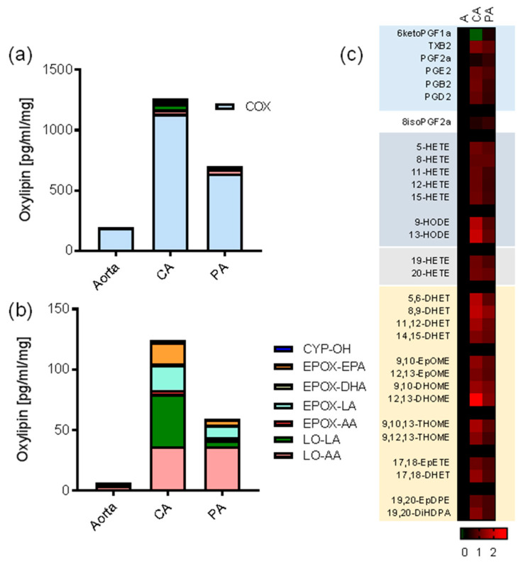
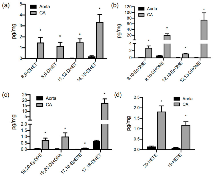
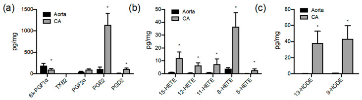
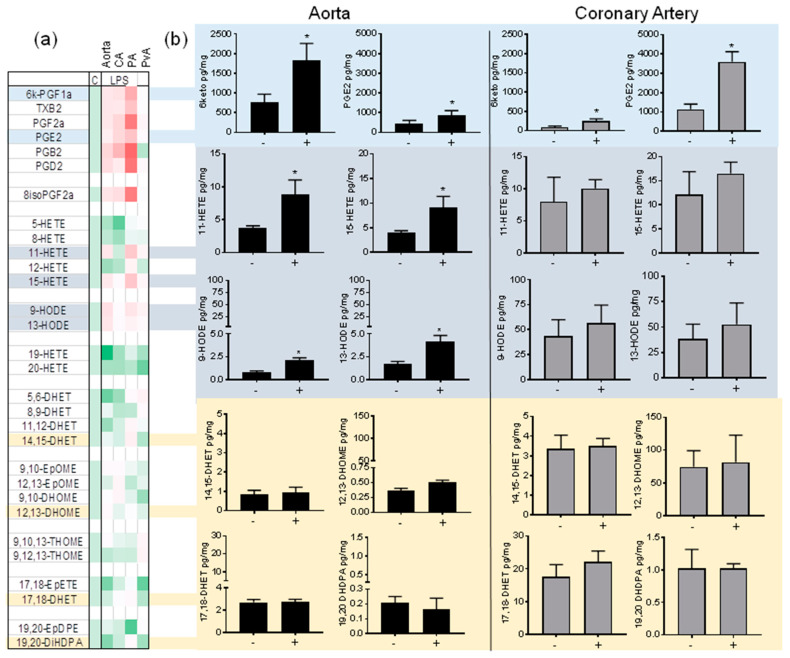
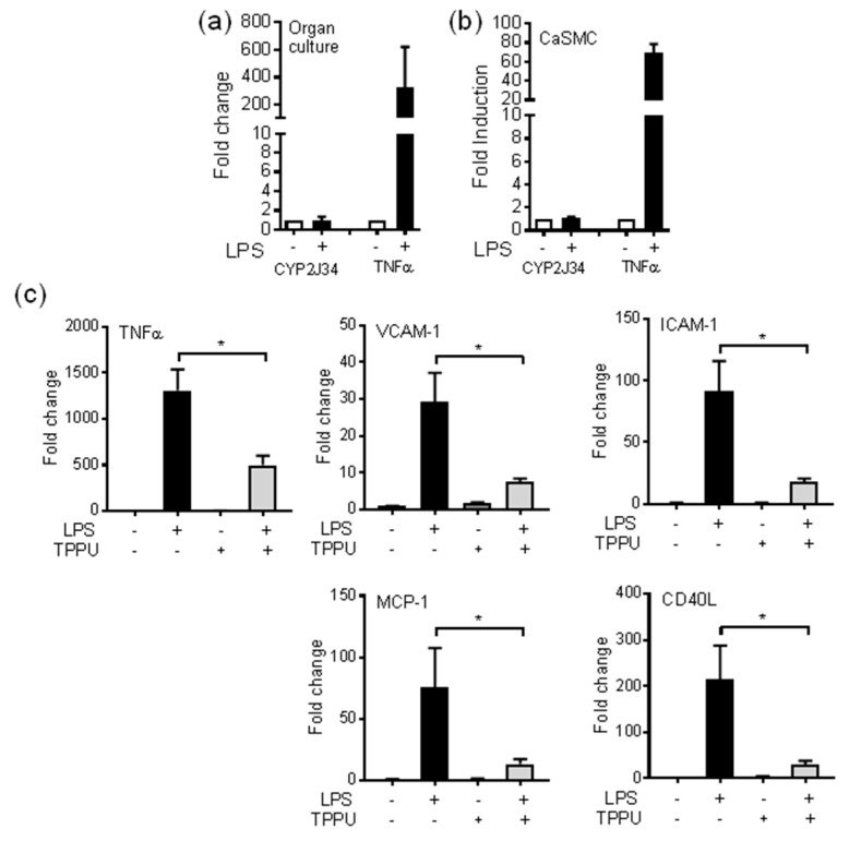
A number of oxylipins have been described as endogenous PPAR ligands. The very short biological half-lives of oxylipins suggest roles as autocrine or paracrine signaling molecules. While coronary arterial atherosclerosis is the root of myocardial infarction, aortic atherosclerotic plaque formation is a common readout of in vivo atherosclerosis studies in mice. Improved understanding of the compartmentalized sources of oxylipin PPAR ligands will increase our knowledge of the roles of PPAR signaling in diverse vascular tissues. Here, we performed a targeted lipidomic analysis of ex vivo-generated oxylipins from porcine aorta, coronary artery, pulmonary artery and perivascular adipose. Cyclooxygenase (COX)-derived prostanoids were the most abundant detectable oxylipin from all tissues. By contrast, the coronary artery produced significantly higher levels of oxylipins from CYP450 pathways than other tissues. The TLR4 ligand LPS induced prostanoid formation in all vascular tissue tested. The 11-HETE, 15-HETE, and 9-HODE were also induced by LPS from the aorta and pulmonary artery but not coronary artery. Epoxy fatty acid (EpFA) formation was largely unaffected by LPS. The pig CYP2J homologue CYP2J34 was expressed in porcine vascular tissue and primary coronary artery smooth muscle cells (pCASMCs) in culture. Treatment of pCASMCs with LPS induced a robust profile of pro-inflammatory target genes: TNFα, ICAM-1, VCAM-1, MCP-1 and CD40L. The soluble epoxide hydrolase inhibitor TPPU, which prevents the breakdown of endogenous CYP-derived EpFAs, significantly suppressed LPS-induced inflammatory target genes. In conclusion, PPAR-activating oxylipins are produced and regulated in a vascular site-specific manner. The CYP450 pathway is highly active in the coronary artery and capable of providing anti-inflammatory oxylipins that prevent processes of inflammatory vascular disease progression.

Keywords: CYP450; PPARs; coronary artery; eicosanoids; inflammation; lipidomics; vascular.


Conflict of interest statement

The authors declare no conflict of interest. The funders had no role in the design of the study; in the collection, analyses, or interpretation of data; in the writing of the manuscript, or in the decision to publish the results.

Figures

Figure 1
Figure 1
Characterization of oxylipin production from aorta, coronary artery, and pulmonary artery. (a,b) Comparative and relative contribution of cyclooxygenase (COX), lipoxygenase-arachidonic acid (LO-AA), lipoxygenase-linoleic acid (LO-LA), CYP-epoxygenase-arachidonic acid (EPOX-AA), CYP-epoxygenase-linoleic acid (EPOX-LA), CYP-epoxygenase-DHA (EPOX-DHA), CYP-epoxygenase-EPA (EPOX-EPA), and CYP-ω-hydroxylase (CYP-OH) products to the oxylipin releasate of aorta, coronary artery (CA), and pulmonary artery (PA) in 24 h organ culture. (a) shows all pathways, whereas (b) shows all pathways minus COX. Bars are based upon the single most oxylipin abundant oxylipin product detected in each pathway which is used as a representative index of oxylipin class. (c) Heatmap showing Log10 fold differences in the mean amount of each oxylipin detected from coronary artery (CA) and pulmonary artery (PA) compared to aorta. The actual fold range in the coronary artery was 0.5-fold for 6-keto PGF to 823-fold for 12,13-DHOME. Data represents organ culture from n = 3–4 separate animals.
Figure 2
Figure 2
Coronary arteries produce high levels of CYP-derived oxylipins. Figures show detectable CYP epoxygenase (a) EPOX-AA, (b) EPOX-LA, (c) EPOX-DHA/EPA and (d) CYP-OH products released by pig aorta (black bars) and coronary artery (grey bars). Oxylipins accumulated in 24 h serum-free organ culture were measured by LC–MS/MS and expressed as pg/mg of wet tissue weight. Data represents organ culture from n = 3–4 separate animals. Data represents organ culture from n = 3–4 separate animals. * indicates p < 0.05 between Aorta and CA.
Figure 3
Figure 3
Comparison of aortic and coronary artery production of cyclooxygenase and ‘lipoxygenase’ oxylipin products. Figures show (a) cyclooxygenase, (b) ‘lipoxygenase’ products of arachidonic acid (HETEs) and (c) linoleic acid (HODEs) products released by pig aorta (black bars) and coronary artery (grey bars). Oxylipins accumulated in 24 h serum-free organ culture were measured by LC–MS/MS and expressed as pg/mg of wet tissue weight. Data represents organ culture from n = 3–4 separate animals. * indicates p < 0.05 between Aorta and CA.
Figure 4
Figure 4
Regulation of oxylipin production in large vessels by LPS/TLR4 activation. (a) Heatmap showing summary of fold differences in the mean oxylipin generation in aorta, coronary artery (CA), pulmonary artery (PA) and perivascular adipose (PvA) untreated tissue (C) compared to tissue treated with LPS (1 μg/mL) ex vivo. The range of fold differences was from 0.5- (19-HETE; Aorta) to 9-fold (PGB2; PVA). (b) Comparison of major oxylipin production: 6-ketoPGF, PGE2, 11-HETE, 15-HETE, 9-HODE, 13-HODE, 14,15-DHET, 12,13-DHOME, 17,18-DHET and 19,20-DHDPA in aorta and coronary artery treated in the absence (-) or presence regulation by LPS (1 μg/mL; +). * indicates p < 0.05 by unpaired t-test between tissue treated in the presence of absence of LPS. Data represents organ culture from n = 3–4 separate animals.
Figure 5
Figure 5
The sEH inhibitor TPPU is anti-inflammatory in coronary artery vascular smooth muscle cells. Expression of TNFα and CYP2J34 mRNA in (a) combined pig coronary and pulmonary artery vessels in organ culture at 24 h (n = 4), and (b) pig primary coronary artery cells at 4 h (CaSMCs) in the presence or absence of LPS (1 μg/mL). mRNA was measured by qRT-PCR and fold levels normalized to 18S. (c) Inflammatory target gene expression of TNFα, VCAM-1, ICAM-1, MCP-1 (CCL2) and CD40 in cultures of pCASMCs in the presence or absence LPS (1 μg/mL; 4 h), and/or sEH inhibitor TPPU (1 μM; given as a 1 h pretreatment before addition of LPS). * indicates p < 0.05 by paired t-test between cells treated with TPPU in the presence of absence of LPS. Data represents mean ± SE from n = 4 cultures from two separate animals.

References

    1. Bishop-Bailey D., Wray J. Peroxisome proliferator-activated receptors: A critical review on endogenous pathways for ligand generation. Prostaglandins Other Lipid Mediat. 2003;71:1–22. doi: 10.1016/S0090-6980(03)00003-0. - DOI - PubMed
    1. Smilowitz J.T., Zivkovic A.M., Wan Y.J., Watkins S.M., Nording M.L., Hammock B.D., German J.B. Nutritional lipidomics: Molecular metabolism, analytics, and diagnostics. Mol. Nutr. Food Res. 2013;57:1319–1335. doi: 10.1002/mnfr.201200808. - DOI - PMC - PubMed
    1. Zeldin D.C. Epoxygenase pathways of arachidonic acid metabolism. J. Biol. Chem. 2001;276:36059–36062. doi: 10.1074/jbc.R100030200. - DOI - PubMed
    1. Bishop-Bailey D., Thomson S., Askari A., Faulkner A., Wheeler-Jones C. Lipid-metabolizing CYPs in the regulation and dysregulation of metabolism. Annu. Rev. Nutr. 2014;34:261–279. doi: 10.1146/annurev-nutr-071813-105747. - DOI - PubMed
    1. Bailey J.M., Bryant R.W., Whiting J., Salata K. Characterization of 11-HETE and 15-HETE, together with prostacyclin, as major products of the cyclooxygenase pathway in cultured rat aorta smooth muscle cells. J. Lipid Res. 1983;24:1419–1428. - PubMed

Publication types

MeSH terms

Substances