Review
. 2020 Apr 29;9(5):1097.
doi: 10.3390/cells9051097.

Towards a Comprehensive Understanding of UA-ADRCs (Uncultured, Autologous, Fresh, Unmodified, Adipose Derived Regenerative Cells, Isolated at Point of Care) in Regenerative Medicine

Affiliations
Review

Towards a Comprehensive Understanding of UA-ADRCs (Uncultured, Autologous, Fresh, Unmodified, Adipose Derived Regenerative Cells, Isolated at Point of Care) in Regenerative Medicine

Eckhard U Alt et al. Cells. .

Abstract

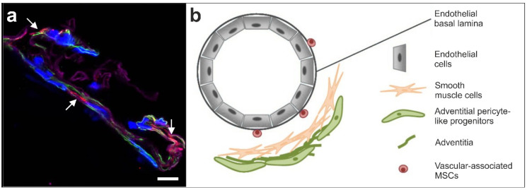
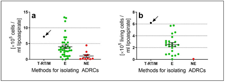
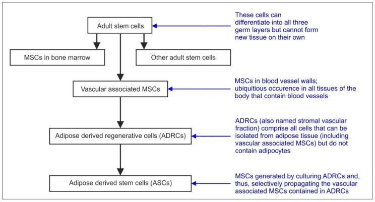
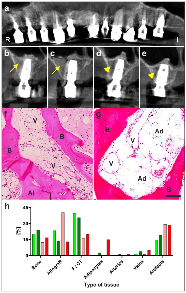
It has become practically impossible to survey the literature on cells derived from adipose tissue for regenerative medicine. The aim of this paper is to provide a comprehensive and translational understanding of the potential of UA-ADRCs (uncultured, unmodified, fresh, autologous adipose derived regenerative cells isolated at the point of care) and its application in regenerative medicine. We provide profound basic and clinical evidence demonstrating that tissue regeneration with UA-ADRCs is safe and effective. ADRCs are neither 'fat stem cells' nor could they exclusively be isolated from adipose tissue. ADRCs contain the same adult stem cells ubiquitously present in the walls of blood vessels that are able to differentiate into cells of all three germ layers. Of note, the specific isolation procedure used has a significant impact on the number and viability of cells and hence on safety and efficacy of UA-ADRCs. Furthermore, there is no need to specifically isolate and separate stem cells from the initial mixture of progenitor and stem cells found in ADRCs. Most importantly, UA-ADRCs have the physiological capacity to adequately regenerate tissue without need for more than minimally manipulating, stimulating and/or (genetically) reprogramming the cells for a broad range of clinical applications. Tissue regeneration with UA-ADRCs fulfills the criteria of homologous use as defined by the regulatory authorities.

Keywords: ADRCs; adipose tissue derived regenerative cells; efficacy; point of care treatment; randomized controlled trials; review; safety; stem cells; stromal vascular fraction.


Conflict of interest statement

E.A. is Executive Chair of InGeneron, Inc. (Houston, TX, USA). G.W. is employed as Chief Scientific Officer at InGeneron, Inc. C.A. is Director Medical and Regulatory Affairs (Munich, Germany) which is owned by InGeneron, Inc. C.S. serves as consultant to InGeneron, Inc. A.H. declares no conflict of interest. InGeneron, Inc. and InGeneron, GmbH had no role in the design of the study; in the collection, analyses, or interpretation of data; in the writing of the manuscript, or in the decision to publish the results.

Figures

Figure 1
Figure 1
(a) Immunofluorescent detection of NG2 (red; arrows), smooth muscle antigen (SMA) (green) and laminin (purple) in the wall of a small arteriole; cell nuclei are in blue (modified from [3]). The scale bar represents 10 µm. (b) Schematic illustration of the hypothesized location of vascular associated MSCs (taken from [3] and modified from [35]).
Figure 2
Figure 2
Comparison of cell yield (a) and number of living cells per mL lipoaspirate (b) of adipose derived regenerative cells (ADRCs) isolated with the Transpose RT/Matrase system (InGeneron) (T-RT/M; black dots) as well as other enzymatic (E; green dots) and non-enzymatic (NE; red dots) methods for isolating ADRCs reported in the literature (modified from [4]). The panels show individual data (dots) as well as mean and standard error of the mean (SEM) in case of the other enzymatic and non-enzymatic methods. One can immediately see that on average enzymatic methods result in a much higher cell yield than non-enzymatic methods. Furthermore, for most of the non-enzymatic methods the number of living cells per mL lipoaspirate could not be calculated because the corresponding relative numbers of living cells were not reported. Of all reported methods the Transpose RT/Matrase system (InGeneron) did not result in the highest cell yield (arrow in a) but in the highest number of living cells per mL lipoaspirate (arrow in b), which appears to be the clinically most relevant parameter (as outlined in the main text).
Figure 3
Figure 3
Schematic overview of the relationship between the term adult stem cells, vascular associated MSCs, ADRCs and ASCs (modified from [3]).
Figure 4
Figure 4
Example of regeneration of bone with UA-ADRCs (modified from [7]). Details are provided in the main text. (ae) digital volume tomography radiographs taken 1 year after the placement of implants and 20 months after guided bone regeneration-maxillary sinus augmentation. Panel a provides a panoramic view; Panels (be) show detailed views on selected regions. With the application of unmodified autologous adipose-derived regenerative cells (UA-ADRCs) the bone around the implants in regions B and C appears larger in area and denser (yellow arrows in (b,c)) than without the application of UA-ADRCs in (d,e) (yellow arrowheads in (d,e)). (f,g) Photomicrographs of bone biopsies taken at six weeks (W6) post treatment with application of cells (f) or without cells (g). (h) Results of histomorphometric analysis of bone biopsies taken at W6 (light green and orange bars) and W34 (dark green and red bars) post treatment with application of cells (green bars) or without cells (orange and red bars). Abbreviations: R, right; L, left; B, bone; Al, allograft; V, vein; Ad, adipocyte; F/CT, fibrin and connective tissue. With cells there was considerably more bone and connective tissue formed already at six weeks than was achieved without cells even after six months. The scale bar in g represents 100 µm.
Figure 5
Figure 5
Example of regeneration of knee cartilage with UA-ADRCs (modified from [4]). The letters (ah) are explained in detail in the main text above. The scale bar in h represents 100 µm.
Figure 6
Figure 6
Representative, previously unpublished example how autologous, adipose derived stem cells can stay locally, survive and engraft in the new host tissue into which the cells were applied, differentiate under guidance of the new microenvironment, integrate into the new host tissue and participate in building new vascular structures in the host tissue. The panels show photomicrographs of paraffin-embedded, 5 µm thick tissue sections of a post mortem heart from a pig, taken from the left ventricular border zone of myocardial infarction ten weeks after experimental occlusion of the left anterior descending (LAD) artery for three hours, followed by delivery of eGFP-labeled autologous ASCs into the balloon-blocked LAD vein (matching the initial LAD occlusion site) at four weeks after occlusion of the LAD (experiments are described in detail in [111]). (ae) One tissue section was stained with DAPI (blue) (a) and processed for immunofluorescent detection of GFP (green) (b), von Willebrand factor (vWF) (red) (c) and Troponin (yellow) (d). The arrows indicate cell nuclei that were immunopositive for GFP and were found in the wall of small vessels (the positions of these cell nuclei are also labeled in the panel representing vWF). (fj) Another tissue section was was stained with DAPI (blue) (f) and processed for immunofluorescent detection of GFP (green) (g), Cx43 (red) (h) and Troponin (yellow) (i). The circles indicate regions where most of the cell nuclei were immunopositive for GFP, and the arrow a GFP-positive cell nucleus inside (or directly adjacent to) a cardiomyocyte. (k–o). A third tissue section was stained with DAPI (blue) (k) and processed for immunofluorescent detection of GFP (green) (l), Ki-67 (red) (m) and Troponin (yellow) (n). The white arrows point to cell nuclei that were immunopositive for GFP but not for Ki-67, the yellow arrows to a cell nucleus that was immunopositive for Ki-67 but not for GFP, and the red arrows to a cell nucleus that was immunopositive for both GFP and Ki-67 (indicating that this cell had re-entered the cell cycle). The scale bar represents 25 µm in the merged panels and 50 µm in the individual panels.

References

    1. Davidoff M.S. The pluripotent microvascular pericytes are the adult stem cells even in the testis. Adv. Exp. Med. Biol. 2019;1122:235–267. - PubMed
    1. Stem Cell Key Terms. [(accessed on 16 April 2020)]; Available online: https://www.cirm.ca.gov/patients/stem-cell-key-terms.
    1. Alt E.U., Schmitz C., Bai X. Fundamentals of stem cells: Why and how patients’ own adult stem cells are the next generation of medicine. In: Trafny T., Spiri S., editors. Bioethics and Research on Adult Stem Cells. IF Press Pontifical Council for Culture; Rome, Italy: 2020. in press.
    1. Winnier G.E., Valenzuela N., Peters-Hall J., Kellner J., Alt C., Alt E.U. Isolation of adipose tissue derived regenerative cells from human subcutaneous tissue with or without the use of an enzymatic reagent. PLoS ONE. 2019;14:e0221457. doi: 10.1371/journal.pone.0221457. - DOI - PMC - PubMed
    1. Crisan M., Yap S., Casteilla L., Chen C.W., Corselli M., Park T.S., Andriolo G., Sun B., Zheng B., Zhang L., et al. A perivascular origin for mesenchymal stem cells in multiple human organs. Cell Stem Cell. 2008;3:301–313. doi: 10.1016/j.stem.2008.07.003. - DOI - PubMed

Publication types