Review
. 2020 Apr 29;21(9):3134.
doi: 10.3390/ijms21093134.

Targeting Mitochondrial Network Architecture in Down Syndrome and Aging

Affiliations
Review

Targeting Mitochondrial Network Architecture in Down Syndrome and Aging

Nunzia Mollo et al. Int J Mol Sci. .

Abstract

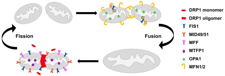
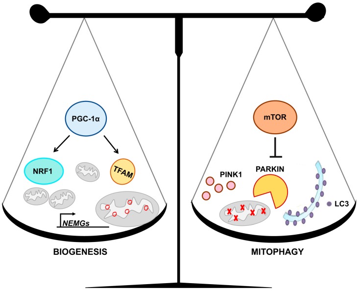
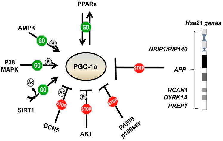
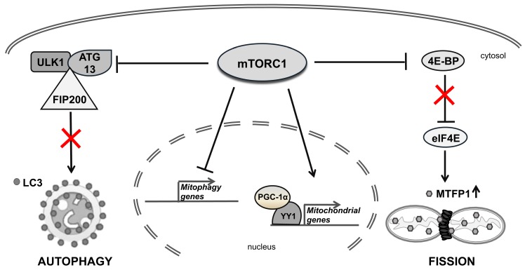
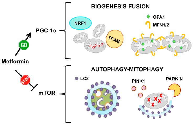
Mitochondria are organelles that mainly control energy conversion in the cell. In addition, they also participate in many relevant activities, such as the regulation of apoptosis and calcium levels, and other metabolic tasks, all closely linked to cell viability. Functionality of mitochondria appears to depend upon their network architecture that may dynamically pass from an interconnected structure with long tubular units, to a fragmented one with short separate fragments. A decline in mitochondrial quality, which presents itself as an altered structural organization and a function of mitochondria, has been observed in Down syndrome (DS), as well as in aging and in age-related pathologies. This review provides a basic overview of mitochondrial dynamics, from fission/fusion mechanisms to mitochondrial homeostasis. Molecular mechanisms determining the disruption of the mitochondrial phenotype in DS and aging are discussed. The impaired activity of the transcriptional co-activator PGC-1α/PPARGC1A and the hyperactivation of the mammalian target of rapamycin (mTOR) kinase are emerging as molecular underlying causes of these mitochondrial alterations. It is, therefore, likely that either stimulating the PGC-1α activity or inhibiting mTOR signaling could reverse mitochondrial dysfunction. Evidence is summarized suggesting that drugs targeting either these pathways or other factors affecting the mitochondrial network may represent therapeutic approaches to improve and/or prevent the effects of altered mitochondrial function. Overall, from all these studies it emerges that the implementation of such strategies may exert protective effects in DS and age-related diseases.

Keywords: Down syndrome; PGC-1α/PPARGC1A; aging; mTOR; mitochondrial dynamics; mitochondrial function; mitochondrial network.


Conflict of interest statement

The authors declare no conflicts of interest.

Figures

Figure 1
Figure 1
The molecular machinery of mitochondrial network regulation. On the left, the mitochondrial fission process in which several proteins are involved. Dynamin-related protein 1 (DRP1), localized in the cytosol, is recruited by fission 1 protein (FIS1), MID49/51, mitochondrial fission factor (MFF) and mitochondrial fission process protein 1 (MTFP1) to the mitochondrial surface, where it forms a membrane constriction ring. On the right, the mitochondrial fusion process that involves optic atrophy gene 1 (OPA1), which mediates fusion of the inner mitochondrial membrane, and mitofusin 1/2 (MFN1/2), which enhance the fusion of the outer mitochondrial membrane. In mammals, FIS1 can act as a fusion machinery inhibitor by binding to OPA1 and MFN1/2 and blocking their activity.
Figure 2
Figure 2
Mitochondrial homeostasis is based on the balanced interplay between biogenesis and mitophagy. On the left, some of the molecular mechanisms of mitochondrial biogenesis are represented. The peroxisome-proliferator-activated receptor γ co-activator-1α (PGC-1α) induces the expression of nuclear respiratory factor 1 (NRF1), which regulates most of the nuclear encoded mitochondrial genes (NEMGs), and transcription factor A (TFAM), which governs mitochondrial DNA (mtDNA). On the right, some aspects of the mitophagy process are represented. Mammalian target of rapamycin (mTOR) signaling negatively regulates the clearance of damaged mitochondria through the pathway dependent upon the PTEN-induced putative kinase 1 (PINK1) and the parkin RBR E3 ubiquitin protein ligase (PARKIN). LC3 promotes autophagosomes formation.
Figure 3
Figure 3
Factors regulating PGC-1α expression and/or activity. The peroxisome-proliferator-activated receptors (PPARs) regulate, and are regulated by, PGC-1α. AMPK, P38 MAPK and SIRT1 positively regulate PGC-1α at post-transcriptional levels. GCN5, AKT, PARIS and p160MBP negatively regulate PGC-1α. PGC-1α expression and/or activity is also regulated by genes mapping to chromosome 21.
Figure 4
Figure 4
The role of mammalian target of rapamycin complex1 (mTORC1) in regulating mitochondrial homeostasis. mTORC1 inhibits autophagy by phosphorylating the regulatory complex formed by unc-51–like kinase (ULK1) and its interacting proteins, autophagy-related protein 13 (ATG13) and focal adhesion kinase family interacting protein of 200 kDa (FIP200) (on the left). mTORC1 stimulates the mitochondrial fission by phosphorylating 4E-BPs, thus promoting translation initiation of MTFP1 (on the right). mTORC1 also represses mitophagy gene expression and regulates the expression of several mitochondrial genes (in the center).
Figure 5
Figure 5
Mitochondrial network and ultrastructure are altered in cells with trisomy of chromosome 21. (A) Representative fluorescence microscopy images showing that the mitochondrial network is fragmented in trisomic cells, which display shorter mitochondria instead of branched elongated tubular ones (higher magnification pictures in the inset). Mitochondria were labelled with a mitochondria-targeted green fluorescent protein. (B) Representative electron microscopy images showing that mitochondria of trisomic cells are damaged and are characterized by a range of alterations: broken, shorter and less numerous cristae (arrow heads). Moreover, giant swollen mitochondria are also observed in trisomic cells (arrows). Scale bar represents 1 μm.
Figure 6
Figure 6
Metformin counteracts mitochondrial network fragmentation in cells with trisomy of chromosome 21. Representative fluorescence microscopy images showing a branched and elongated tubular morphology of the mitochondrial network in trisomic cells treated with metformin at two different concentrations. Mitochondria were labelled with a mitochondria-targeted green fluorescent protein.
Figure 7
Figure 7
Metformin action on mitochondrial homeostasis. Metformin acts on mitochondrial homeostasis by two pathways: on one side it increases mitochondrial biogenesis and induces mitochondrial fusion via PGC-1α activation. On the other side it induces the formation of autophagosomes and promotes the clearance of damaged mitochondria via mTOR inhibition.

References

    1. Spinelli J.B., Haigis M.C. The multifaceted contributions of mitochondria to cellular metabolism. Nat. Cell Biol. 2018;20:745–754. doi: 10.1038/s41556-018-0124-1. - DOI - PMC - PubMed
    1. Duchen M.R. Mitochondria and calcium: From cell signalling to cell death. J. Physiol. 2000;529:57–68. doi: 10.1111/j.1469-7793.2000.00057.x. - DOI - PMC - PubMed
    1. Wang C., Youle R.J. The role of mitochondria in apoptosis*. Ann. Rev. Genet. 2009;43:95–118. doi: 10.1146/annurev-genet-102108-134850. - DOI - PMC - PubMed
    1. Maeda Y., Chida J. Control of cell differentiation by mitochondria, typically evidenced in dictyostelium development. Biomolecules. 2013;3:943–966. doi: 10.3390/biom3040943. - DOI - PMC - PubMed
    1. Buck M.D., O’Sullivan D., Klein Geltink R.I., Curtis J.D., Chang C.H., Sanin D.E., Qiu J., Kretz O., Braas D., van der Windt G.J., et al. Mitochondrial Dynamics Controls T Cell Fate through Metabolic Programming. Cell. 2016;166:63–76. doi: 10.1016/j.cell.2016.05.035. - DOI - PMC - PubMed