. 2020 Apr 30;21(9):3154.
doi: 10.3390/ijms21093154.

HIV-1-Infected Human Macrophages, by Secreting RANK-L, Contribute to Enhanced Osteoclast Recruitment

Affiliations

HIV-1-Infected Human Macrophages, by Secreting RANK-L, Contribute to Enhanced Osteoclast Recruitment

Rémi Mascarau et al. Int J Mol Sci. .

Abstract

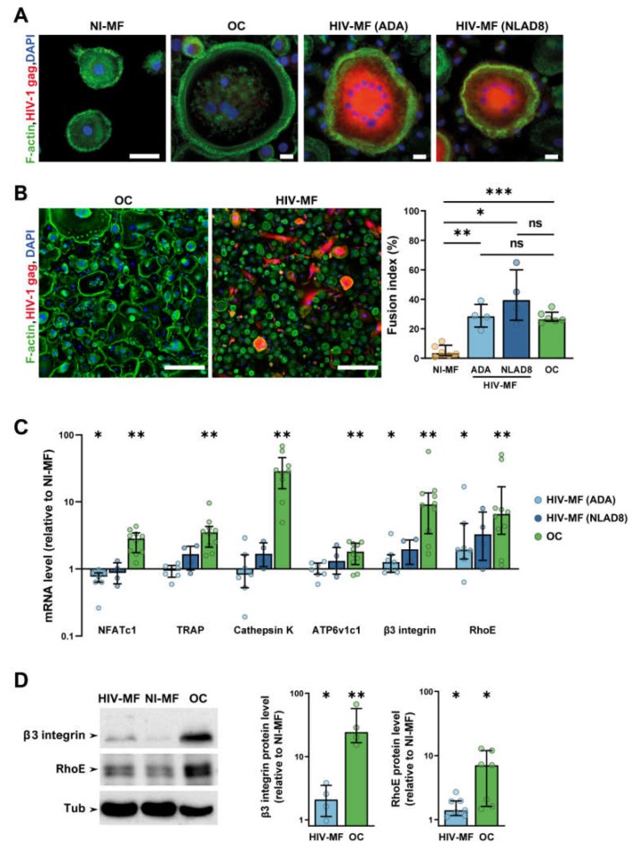
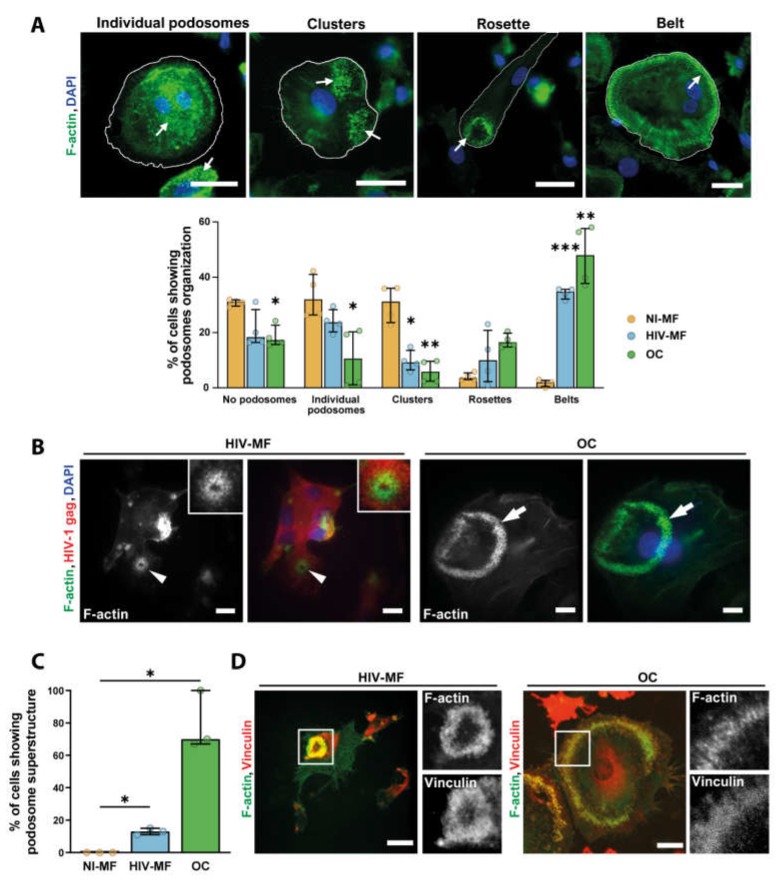
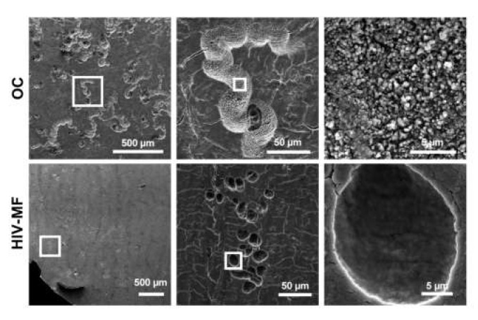
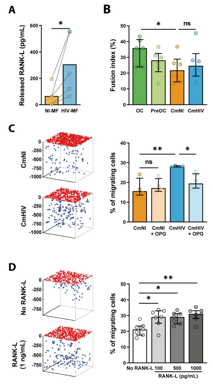
HIV-1 infection is frequently associated with low bone density, which can progress to osteoporosis leading to a high risk of fractures. Only a few mechanisms have been proposed to explain the enhanced osteolysis in the context of HIV-1 infection. As macrophages are involved in bone homeostasis and are critical host cells for HIV-1, we asked whether HIV-1-infected macrophages could participate in bone degradation. Upon infection, human macrophages acquired some osteoclast features: they became multinucleated, upregulated the osteoclast markers RhoE and β3 integrin, and organized their podosomes as ring superstructures resembling osteoclast sealing zones. However, HIV-1-infected macrophages were not fully differentiated in osteoclasts as they did not upregulate NFATc-1 transcription factor and were unable to degrade bone. Investigating whether infected macrophages participate indirectly to virus-induced osteolysis, we showed that they produce RANK-L, the key osteoclastogenic cytokine. RANK-L secreted by HIV-1-infected macrophages was not sufficient to stimulate multinucleation, but promoted the protease-dependent migration of osteoclast precursors. In conclusion, we propose that, by stimulating RANK-L secretion, HIV-1-infected macrophages contribute to create a microenvironment that favors the recruitment of osteoclasts, participating in bone disorders observed in HIV-1 infected patients.

Keywords: HIV-1; RANK-L; bone defects; cell migration; macrophages; osteoclasts.


Conflict of interest statement

The authors have declared that no conflict of interest exists.

Figures

Figure 1
Figure 1
HIV-1 infection induces macrophage (MF) fusion and the expression of some osteoclast (OC) markers. MF were infected or not with HIV-1 (indicated strains), and compared to autologous OC for cell fusion (A,B) and expression of different OC markers (C,D). (A) Representative immunofluorescence (IF) images of uninfected MF (NI-MF), MF infected with HIV-1 (HIV-MF, ADA, or NLAD8 strain), and OC after staining for HIV-1 gag (red), F-actin (green), and nuclei (DAPI, blue). Scale bar, 10 µm. (B) Left panels: Representative large IF images of HIV-MF (ADA strain) and OC after staining for F-actin (green), HIV-1 gag (red), and nuclei (DAPI, blue). Scale bar, 200 µm. Right panel: Quantification of the fusion index evaluated by IF, corresponding to the percentage of nuclei within multinucleated cells. Histograms represent median and error bars are interquartile range, n = 4 to 6 donors, 300 cells analyzed per donor and per condition. (C) mRNA expression of genes overexpressed in OC measured by RT-PCR using the ΔΔCT method in HIV-MF (ADA or NLAD8 strain) and in OC. Actin mRNA level was used as control. Values are normalized to mRNA level in autologous uninfected MF. Histograms represent median and error bars are interquartile range, n = 4 to 10 donors. (D) NI-MF, HIV-MF (NLAD8 strain), and OC lysates were subjected to Western blot using antibodies against β3-integrin, RhoE, and α-Tubulin as loading control. A representative blot (left panel) and quantification of the protein level ratio over autologous NI-MF (right panel) are shown. Histograms represent median and error bars are interquartile range, n = 4 to 7 donors. * p ≤ 0.05; ** p ≤ 0.01; *** p ≤ 0.001, ns: not significantly different.
Figure 2
Figure 2
HIV-1 infection of MF promotes podosome organization into super-structures. (A) Top: Representative IF images of different podosome organizations in MF or OC seeded on glass coverslips, after staining for F-actin (green) and nuclei (DAPI, blue). White arrows show podosome structures. Scale bar, 20 µm. Bottom: Quantification of the different podosome organizations in non-infected MF (NI-MF), HIV-1-infected MF (HIV-MF, ADA strain), and OC represented in percentage of total cells. Histograms represent median and error bars are in interquartile range, n = 4 donors, 300 cells analyzed per donor. (B) Monocytes were seeded on bone slices and differentiated into MF or OC. Then, at day 7, MF were infected with HIV-1 (NLAD8 strain) and all cells were fixed at day 14. Left: representative IF images of podosome super structures in HIV-MF (arrowheads) and OC (arrows) stained for HIV-1 gag (red), F-actin (green), and nuclei (DAPI, blue). Scale bar, 5 µm. Inserts show two-fold magnification of the podosome structure in HIV-MF. (C) Quantification of the percentage of MGC showing podosome organization on bones, evaluated by IF. Histograms represent median and error bars are in interquartile range, n = 3 donors, 100 cells analyzed per condition. (D) Representative image of podosome super-structures formed by HIV-MF (NLAD8 strain, left) or OC (right) seeded on bone slices, after staining for F-actin (green) and vinculin (red). Scale bar, 10 µm. Inserts show two-fold magnification of white square. * p ≤ 0.05; ** p ≤ 0.01; *** p ≤ 0.001.
Figure 3
Figure 3
Superficial bone resorption by HIV-1-infected MF compared to OC. Monocytes were seeded on bone slices and differentiated into MF or OC. At day 7, MF were infected with HIV-1. At day 14, cells were removed and bone slices were stained with toluidine blue. Representative scanning electron microscopy images showing bone resorption pits formed by OC or infected MF (HIV-MF, NLAD8 strain). Scale bars indicated.
Figure 4
Figure 4
RANK-L is secreted by HIV-1-infected MF and promotes 3D migration of OC precursors. (A) MF were infected with HIV-1 (HIV-MF, NLAD8 strain) for 7 days, and the level of released RANK-L was measured by ELISA in the supernatant and compared to uninfected MF (NI-MF) from the same donor. n = 5. (B) Monocytes were differentiated for 3 days in presence of sub-optimal concentrations of RANK-L and then exposed to the supernatant of infected (CmHIV) or uninfected (CmNI) MF for additional 10 days. Cells were then fixed and the fusion index was quantified by IF. Bars represent median, n = 6 donors. (C, D) CmHIV or CmNI pre-treated or not with OPG (100ng/mL) (C), or different concentrations of recombinant RANK-L (D) was added at the bottom of 3D Matrigel matrices. OC precursors were then seeded at the top of the matrices and allowed to migrate. Left: 3D representation of the positions of OC precursors that have migrated (blue dots) or not (red dots) in a representative migration assay. Ticks represent depth in microns. Right: quantification of the percentage of cells inside the matrix. (C) n = 4 donors; (D) n = 7 donors, performed in triplicates. Histograms represent median and error bars are interquartile range. * p ≤ 0.05; ** p ≤ 0.001, ns: not significantly different.

References

    1. Cotter A.G., Mallon P.W. The effects of untreated and treated HIV infection on bone disease. Curr. Opin. HIV AIDS. 2014;9:17–26. doi: 10.1097/COH.0000000000000028. - DOI - PubMed
    1. Moran C.A., Weitzmann M.N., Ofotokun I. Bone Loss in HIV Infection. Curr. Treat. Options Infect. Dis. 2017;9:52–67. doi: 10.1007/s40506-017-0109-9. - DOI - PMC - PubMed
    1. Bruera D., Luna N., David D.O., Bergoglio L.M., Zamudio J. Decreased bone mineral density in HIV-infected patients is independent of antiretroviral therapy. Aids. 2003;17:1917–1923. doi: 10.1097/00002030-200309050-00010. - DOI - PubMed
    1. Gibellini D., Borderi M., De Crignis E., Cicola R., Vescini F., Caudarella R., Chiodo F., Re M.C. RANK-L/OPG/TRAIL plasma levels and bone mass loss evaluation in antiretroviral naive HIV-1-positive men. J. Med. Virol. 2007;79:1446–1454. doi: 10.1002/jmv.20938. - DOI - PubMed
    1. Grijsen M.L., Vrouenraets S.M., Steingrover R., Lips P., Reiss P., Wit F.W., Prins J.M. High prevalence of reduced bone mineral density in primary HIV-1-infected men. Aids. 2010;24:2233–2238. doi: 10.1097/QAD.0b013e32833c93fe. - DOI - PubMed